Deposition Date
2021-06-28
Release Date
2022-07-20
Last Version Date
2023-10-18
Entry Detail
PDB ID:
7R97
Keywords:
Title:
Crystal structure of postcleavge complex of Escherichia coli RNase III
Biological Source:
Source Organism(s):
Escherichia coli (strain K12) (Taxon ID: 83333)
Escherichia coli (Taxon ID: 562)
Escherichia coli (Taxon ID: 562)
Expression System(s):
Method Details:
Experimental Method:
Resolution:
1.80 Å
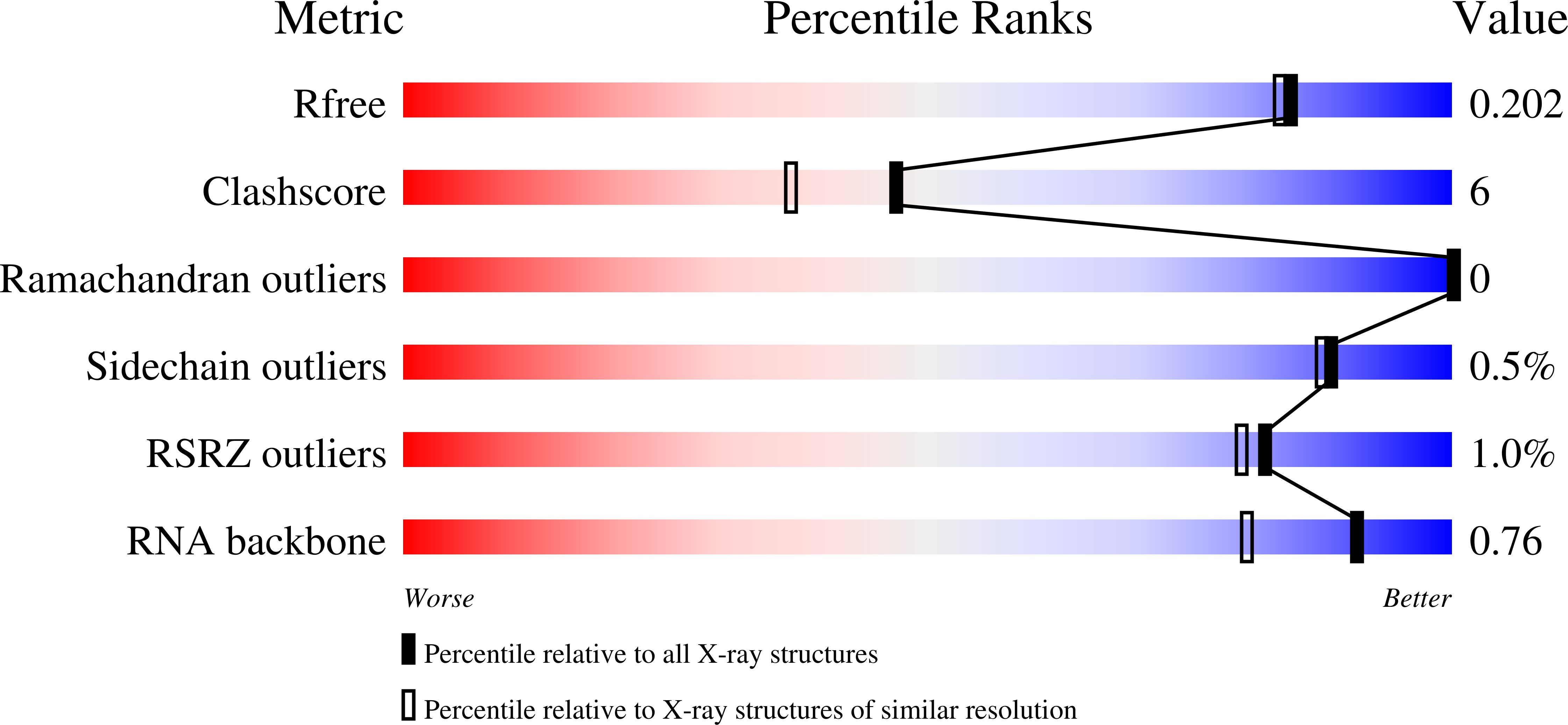
R-Value Free:
0.20
R-Value Work:
0.17
R-Value Observed:
0.17
Space Group:
P 1 21 1