7R1V image
Deposition Date 2022-02-03
Release Date 2022-09-28
Last Version Date 2024-04-03
Entry Detail
PDB ID:
7R1V
Title:
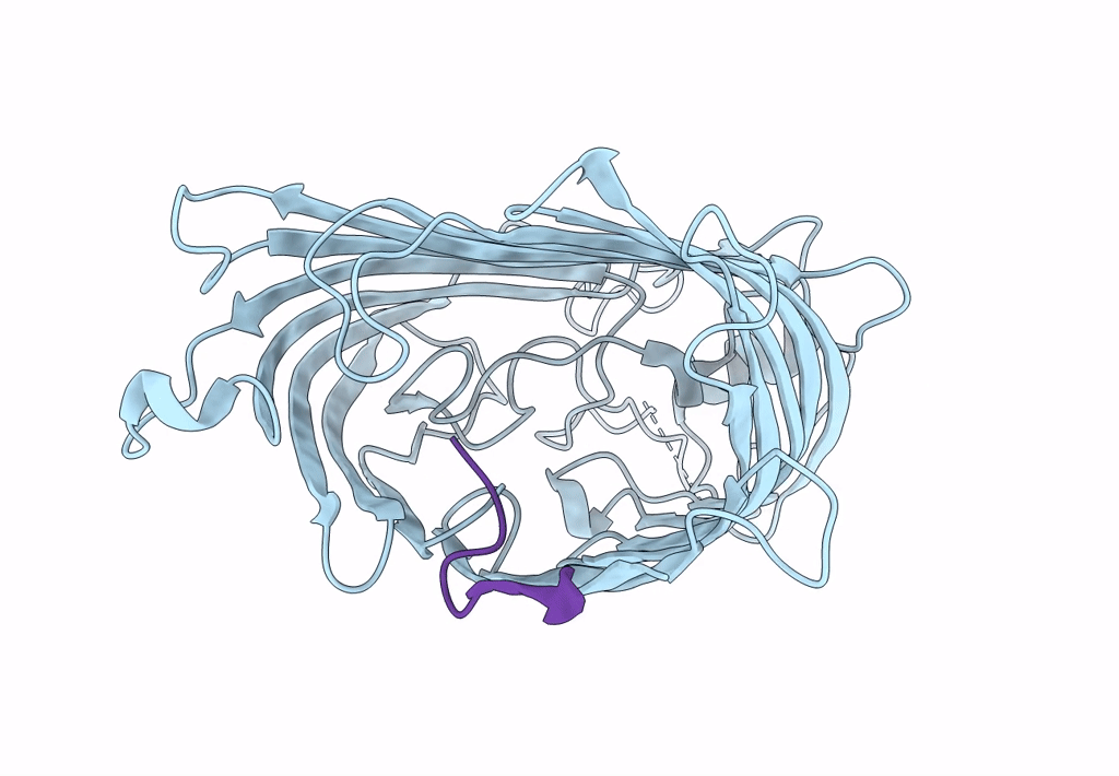
Crystal structure of E.coli BamA beta-barrel in complex with dynobactin A
Biological Source:
Source Organism:
Method Details:
Experimental Method:
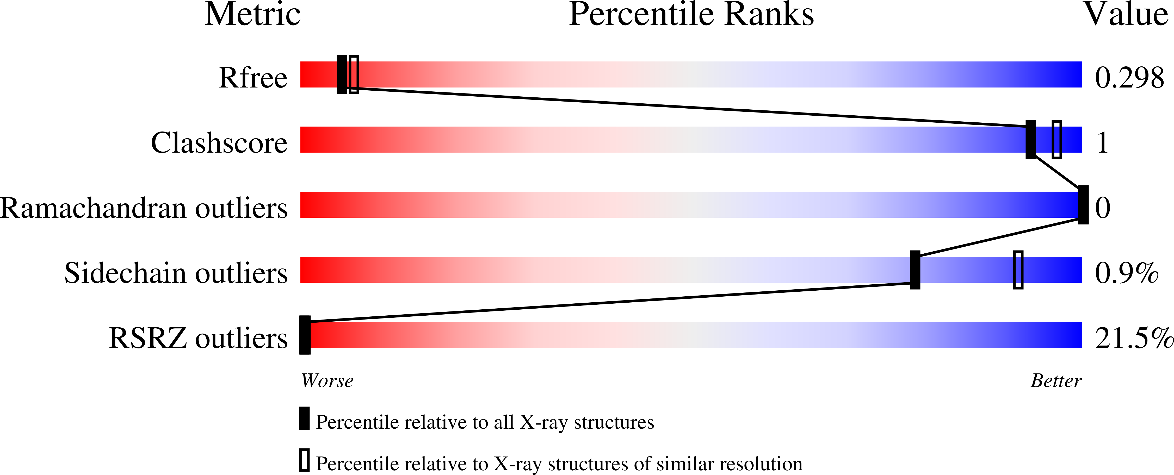
Resolution:
2.50 Å
R-Value Free:
0.27
R-Value Work:
0.24
R-Value Observed:
0.24
Space Group:
P 21 21 21
Macromolecular Entities
Polymer Type:polypeptide(L)
Molecule:Outer membrane protein assembly factor BamA
Chain IDs:A
Chain Length:392
Number of Molecules:1
Biological Source:Escherichia coli O157:H7
Polymer Type:polypeptide(L)
Molecule:Dynobactin A
Gene (Uniprot):Ppb6_00877
Chain IDs:B
Chain Length:10
Number of Molecules:1
Biological Source:Photorhabdus australis
Peptide-like Molecules
PRD_002404
Primary Citation

Abstact

Discovery of antibiotics acting against Gram-negative species is uniquely challenging due to their restrictive penetration barrier. BamA, which inserts proteins into the outer membrane, is an attractive target due to its surface location. Darobactins produced by Photorhabdus, a nematode gut microbiome symbiont, target BamA. We reasoned that a computational search for genes only distantly related to the darobactin operon may lead to novel compounds. Following this clue, we identified dynobactin A, a novel peptide antibiotic from Photorhabdus australis containing two unlinked rings. Dynobactin is structurally unrelated to darobactins, but also targets BamA. Based on a BamA-dynobactin co-crystal structure and a BAM-complex-dynobactin cryo-EM structure, we show that dynobactin binds to the BamA lateral gate, uniquely protruding into its β-barrel lumen. Dynobactin showed efficacy in a mouse systemic Escherichia coli infection. This study demonstrates the utility of computational approaches to antibiotic discovery and suggests that dynobactin is a promising lead for drug development.

Legend

Protein

Chemical

Disease

Primary Citation of related structures