7QZ2 image
Deposition Date 2022-01-30
Release Date 2022-07-06
Last Version Date 2024-02-07
Entry Detail
PDB ID:
7QZ2
Title:
Crystal structure of GacS D1 domain in complex with BeF3-
Biological Source:
Source Organism:
Host Organism:
Method Details:
Experimental Method:
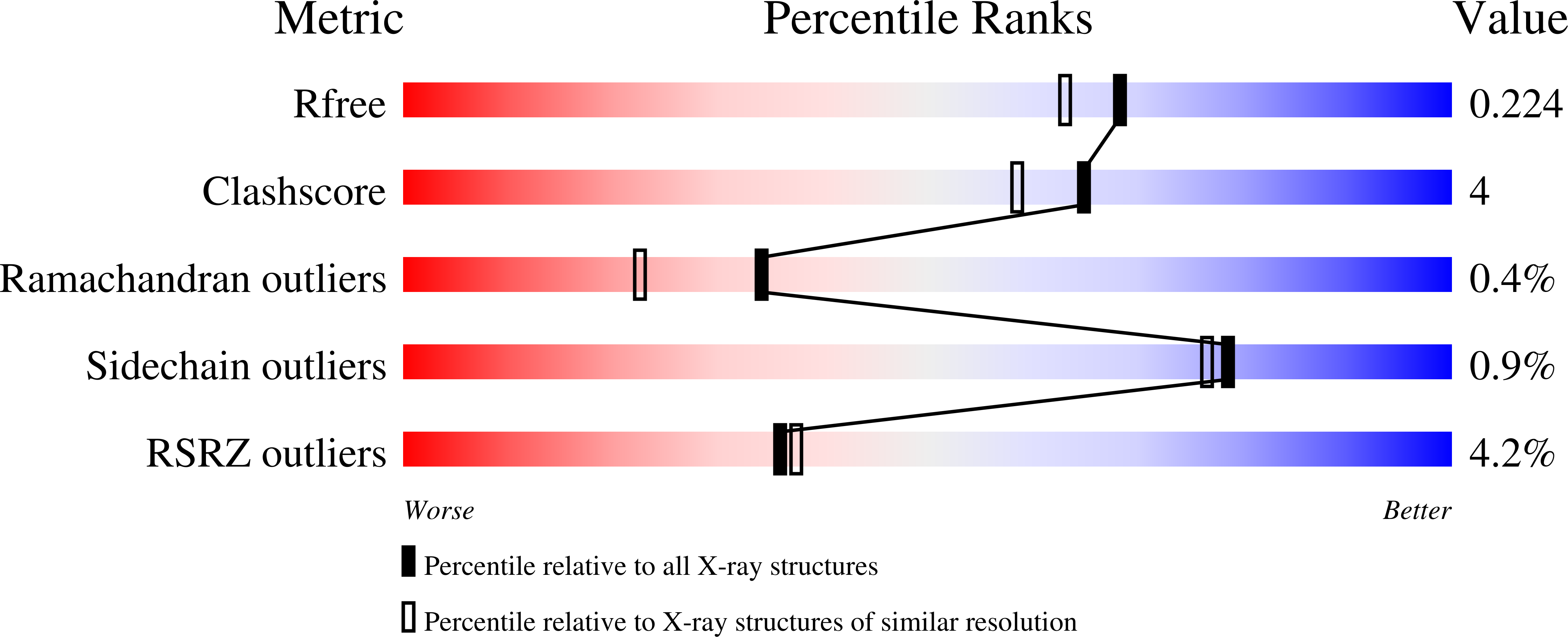
Resolution:
1.87 Å
R-Value Free:
0.21
R-Value Work:
0.16
R-Value Observed:
0.17
Space Group:
C 2 2 21
Macromolecular Entities
Polymer Type:polypeptide(L)
Molecule:Histidine kinase
Chain IDs:A, B
Chain Length:156
Number of Molecules:2
Biological Source:Pseudomonas aeruginosa
Primary Citation
Insights into the atypical autokinase activity of the Pseudomonas aeruginosa GacS histidine kinase and its interaction with RetS.
Structure 30 1285 1297.e5 (2022)
PMID: 35767996 DOI: 10.1016/j.str.2022.06.002

Abstact

Virulence in Pseudomonas aeruginosa (PA) depends on complex regulatory networks, involving phosphorelay systems based on two-component systems (TCSs). The GacS/GacA TCS is a master regulator of biofilm formation, swarming motility, and virulence. GacS is a membrane-associated unorthodox histidine kinase (HK) whose phosphorelay signaling pathway is inhibited by the RetS hybrid HK. Here we provide structural and functional insights into the interaction of GacS with RetS. The structure of the GacS-HAMP-H1 cytoplasmic regions reveals an unusually elongated homodimer marked by a 135 Å long helical bundle formed by the HAMP, the signaling helix (S helix) and the DHp subdomain. The HAMP and S helix regions are essential for GacS signaling and contribute to the GacS/RetS binding interface. The structure of the GacS D1 domain together with the discovery of an unidentified functional ND domain, essential for GacS full autokinase activity, unveils signature motifs in GacS required for its atypical autokinase mechanism.

Legend

Protein

Chemical

Disease

Primary Citation of related structures