7QUT image
Deposition Date 2022-01-18
Release Date 2022-04-13
Last Version Date 2024-01-31
Entry Detail
PDB ID:
7QUT
Title:
serial synchrotron crystallographic structure of Drosophila Melanogaster (6-4) photolyase
Biological Source:
Source Organism(s):
Expression System(s):
Method Details:
Experimental Method:
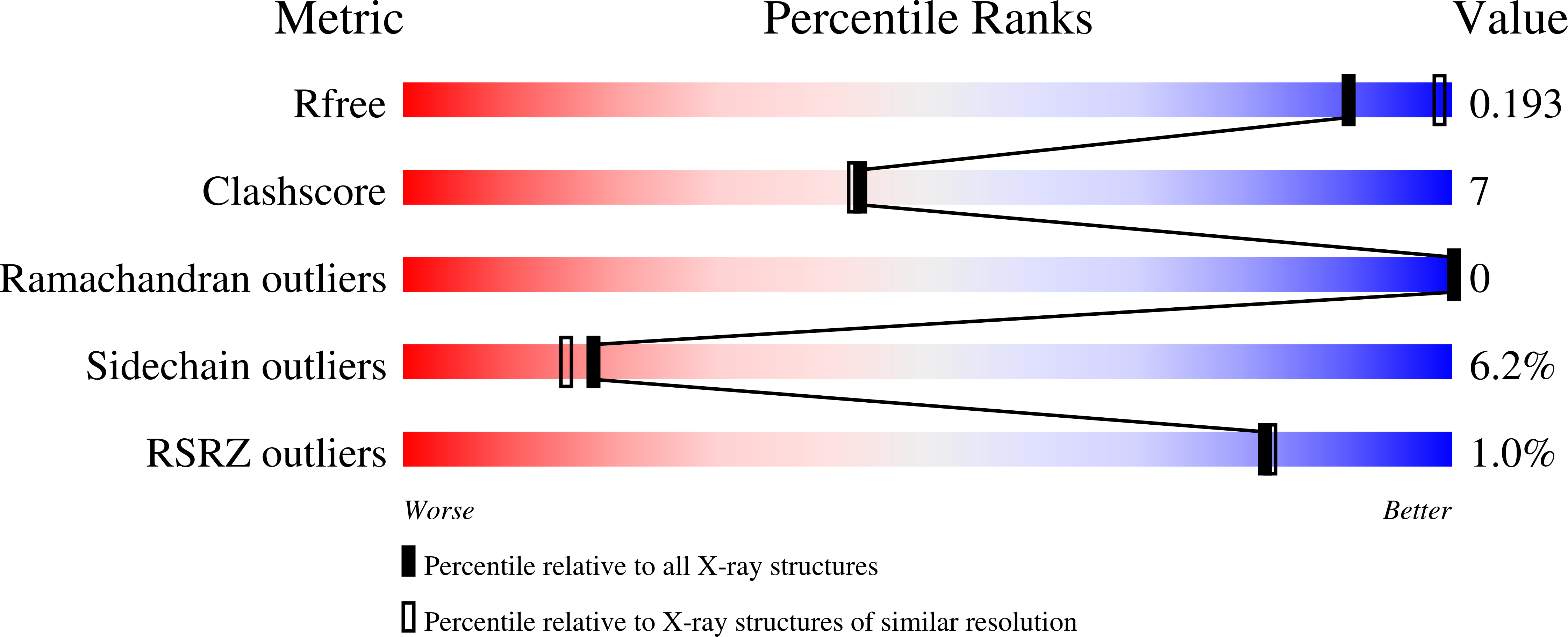
Resolution:
2.24 Å
R-Value Free:
0.19
R-Value Work:
0.15
R-Value Observed:
0.15
Space Group:
P 41
Macromolecular Entities
Structures with similar UniProt ID
Protein Blast
Polymer Type:polypeptide(L)
Molecule:RE11660p
Chain IDs:A
Chain Length:508
Number of Molecules:1
Biological Source:Drosophila melanogaster
Primary Citation
Structural basis of the radical pair state in photolyases and cryptochromes.
Chem.Commun.(Camb.) 58 4889 4892 (2022)
PMID: 35352724 DOI: 10.1039/d2cc00376g

Abstact

We present the structure of a photoactivated animal (6-4) photolyase in its radical pair state, captured by serial crystallography. We observe how a conserved asparigine moves towards the semiquinone FAD chromophore and stabilizes it by hydrogen bonding. Several amino acids around the final tryptophan radical rearrange, opening it up to the solvent. The structure explains how the protein environment stabilizes the radical pair state, which is crucial for function of (6-4) photolyases and cryptochromes.

Legend

Protein

Chemical

Disease

Primary Citation of related structures
Feedback Form
Name
Email
Institute
Feedback