7QUR image
Entry Detail
PDB ID:
7QUR
EMDB ID:
Keywords:
Title:
SARS-CoV-2 Spike with ethylbenzamide-tri-iodo Siallyllactose, C3 symmetry
Biological Source:
PDB Version:
Deposition Date:
2022-01-18
Release Date:
2022-06-01
Method Details:
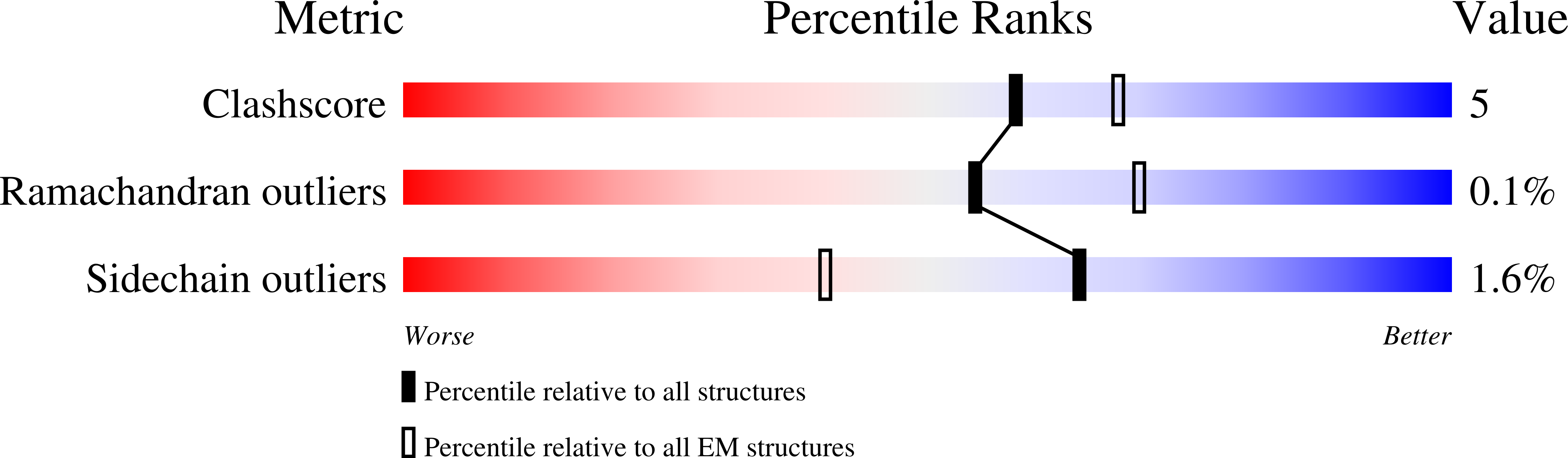
Experimental Method:
Resolution:
2.27 Å
Aggregation State:
PARTICLE
Reconstruction Method:
SINGLE PARTICLE
Macromolecular Entities
Polymer Type:polypeptide(L)
Description:Spike glycoprotein,Fibritin
Chain IDs:A, B, C
Chain Length:1259
Number of Molecules:3
Biological Source:Severe acute respiratory syndrome coronavirus 2, Enterobacteria phage T4
Primary Citation

Abstact

Many pathogens exploit host cell-surface glycans. However, precise analyses of glycan ligands binding with heavily modified pathogen proteins can be confounded by overlapping sugar signals and/or compounded with known experimental constraints. Universal saturation transfer analysis (uSTA) builds on existing nuclear magnetic resonance spectroscopy to provide an automated workflow for quantitating protein-ligand interactions. uSTA reveals that early-pandemic, B-origin-lineage severe acute respiratory syndrome coronavirus 2 (SARS-CoV-2) spike trimer binds sialoside sugars in an "end-on" manner. uSTA-guided modeling and a high-resolution cryo-electron microscopy structure implicate the spike N-terminal domain (NTD) and confirm end-on binding. This finding rationalizes the effect of NTD mutations that abolish sugar binding in SARS-CoV-2 variants of concern. Together with genetic variance analyses in early pandemic patient cohorts, this binding implicates a sialylated polylactosamine motif found on tetraantennary N-linked glycoproteins deep in the human lung as potentially relevant to virulence and/or zoonosis.

Legend

Protein

Chemical

Disease

Primary Citation of related structures