7QPU image
Deposition Date 2022-01-05
Release Date 2022-03-09
Last Version Date 2024-01-31
Entry Detail
PDB ID:
7QPU
Keywords:
Title:
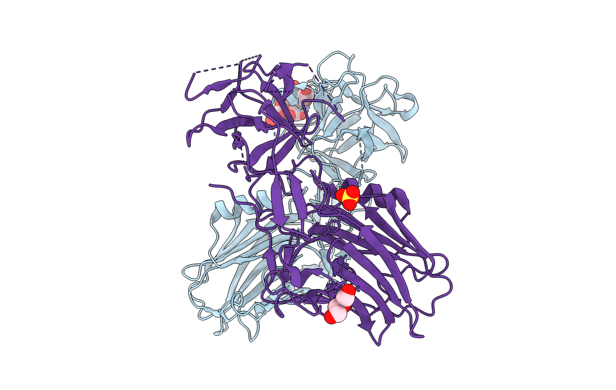
Botulinum neurotoxin A5 cell binding domain in complex with GM1b oligosaccharide
Biological Source:
Source Organism:
Method Details:
Experimental Method:
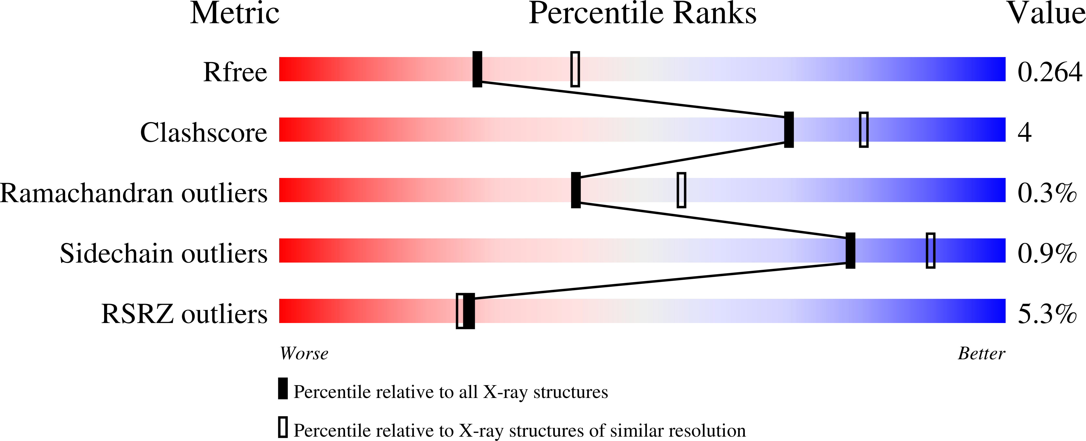
Resolution:
2.40 Å
R-Value Free:
0.26
R-Value Work:
0.21
R-Value Observed:
0.22
Space Group:
P 1 21 1
Macromolecular Entities
Structures with similar UniProt ID
Protein Blast
Polymer Type:polypeptide(L)
Molecule:Botulinum neurotoxin sub-type A5
Gene (Uniprot):cntA
Chain IDs:A, B
Chain Length:433
Number of Molecules:2
Biological Source:Clostridium botulinum
Primary Citation
Crystal Structures of Botulinum Neurotoxin Subtypes A4 and A5 Cell Binding Domains in Complex with Receptor Ganglioside.
Toxins 14 ? ? (2022)
PMID: 35202156 DOI: 10.3390/toxins14020129

Abstact

Botulinum neurotoxins (BoNT) cause the potentially fatal neuroparalytic disease botulism that arises due to proteolysis of a SNARE protein. Each BoNT is comprised of three domains: a cell binding domain (HC), a translocation domain (HN), and a catalytic (Zn2+ endopeptidase) domain (LC). The HC is responsible for neuronal specificity by targeting both a protein and ganglioside receptor at the neuromuscular junction. Although highly toxic, some BoNTs are commercially available as therapeutics for the treatment of a range of neuromuscular conditions. Here we present the crystal structures of two BoNT cell binding domains, HC/A4 and HC/A5, in a complex with the oligosaccharide of ganglioside, GD1a and GM1b, respectively. These structures, along with a detailed comparison with the previously reported apo-structures, reveal the conformational changes that occur upon ganglioside binding and the interactions involved.

Legend

Protein

Chemical

Disease

Primary Citation of related structures