Deposition Date
2022-01-05
Release Date
2022-03-16
Last Version Date
2024-11-06
Entry Detail
PDB ID:
7QPT
Keywords:
Title:
Botulinum neurotoxin A4 cell binding domain in complex with GD1a oligosaccharide
Biological Source:
Source Organism(s):
Clostridium botulinum (Taxon ID: 1491)
Expression System(s):
Method Details:
Experimental Method:
Resolution:
2.30 Å
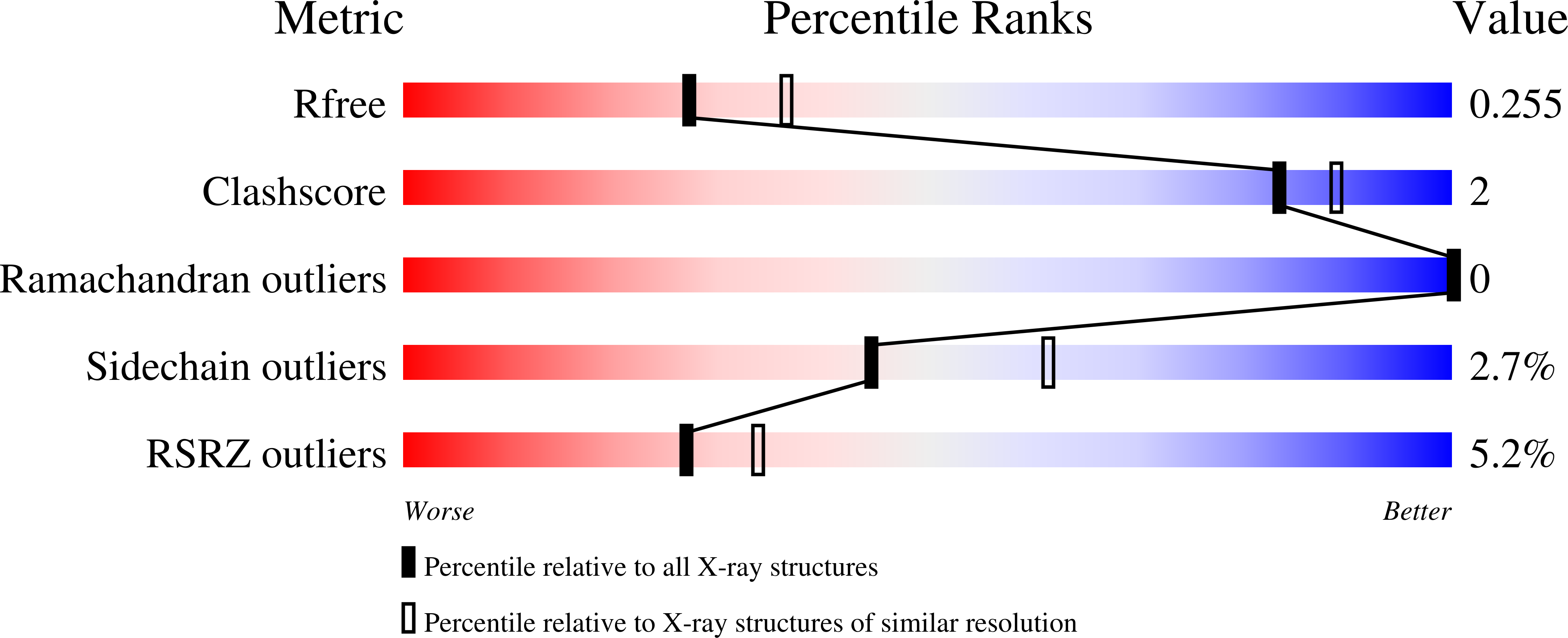
R-Value Free:
0.24
R-Value Work:
0.20
R-Value Observed:
0.20
Space Group:
P 61