7QH3 image
Entry Detail
PDB ID:
7QH3
Keywords:
Title:
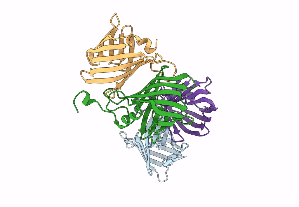
Crystal structure of the anti-sigma factor RsfG from Streptomyces tsukubaensis NRRL18488
Biological Source:
Host Organism:
PDB Version:
Deposition Date:
2021-12-10
Release Date:
2022-12-21
Method Details:
Experimental Method:
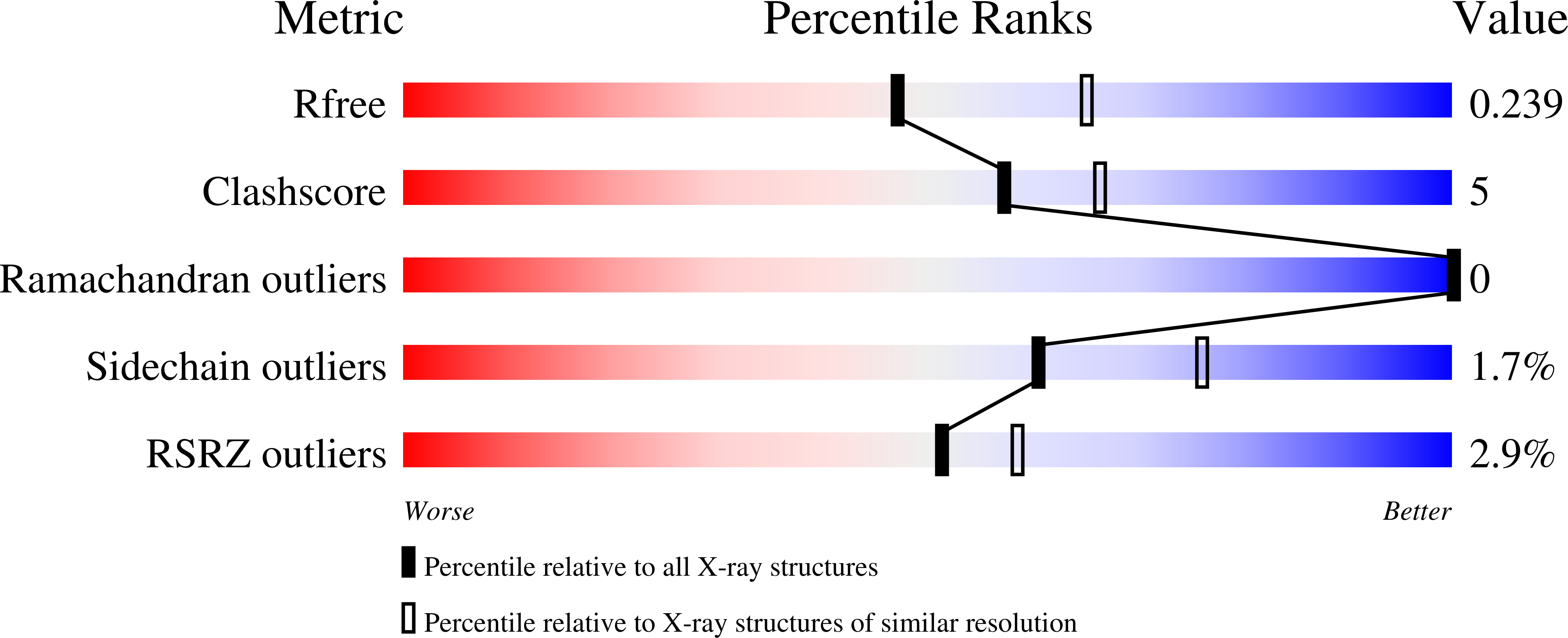
Resolution:
2.30 Å
R-Value Free:
0.24
R-Value Work:
0.19
R-Value Observed:
0.19
Space Group:
P 1 21 1
Macromolecular Entities
Polymer Type:polypeptide(L)
Description:RsfG
Chain IDs:A, B, C, D
Chain Length:154
Number of Molecules:4
Biological Source:Streptomyces tsukubensis NRRL18488
Primary Citation
Crystal structures of Streptomyces tsukubaensis sigma factor SigG1 and anti-sigma RsfG.
J.Struct.Biol. 215 108038 108038 (2023)
PMID: 37858875 DOI: 10.1016/j.jsb.2023.108038

Abstact

Transcription of specific genes in bacteria under environmental stress is frequently initiated by extracytoplasmic function (ECF) σ factors. ECFs σ factors harbour two conserved domains, σ2 and σ4, for transcription initiation by recognition of the promoter region and recruitment of RNA polymerase (RNAP). The crystal structure of Streptomyces tsukubaensis SigG1, an ECF56-family σ factor, was determined revealing σ2, σ4 and the additional carboxi-terminal domain SnoaL_2 tightly packed in a compact conformation. The structure of anti-sigma RsfG was also determined by X-ray crystallography and shows a rare β-barrel fold. Analysis of the metal binding motifs inside the protein barrel are consistent with Fe(III) binding, which is in agreement with previous findings that the Streptomyces tsukubaensis ECF56 SigG1-RsfG system is involved in metal-ion homeostasis.

Legend

Protein

Chemical

Disease

Primary Citation of related structures