7QGK image
Entry Detail
PDB ID:
7QGK
Title:
The mRubyFT protein, Genetically Encoded Blue-to-Red Fluorescent Timer in its red state
Biological Source:
Source Organism:
Host Organism:
PDB Version:
Deposition Date:
2021-12-08
Release Date:
2022-03-23
Method Details:
Experimental Method:
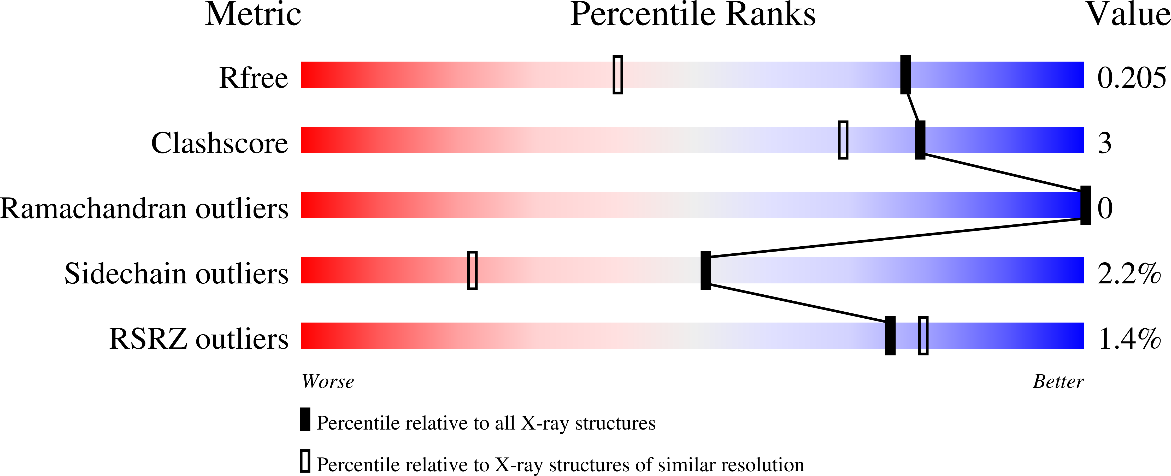
Resolution:
1.50 Å
R-Value Free:
0.19
R-Value Work:
0.17
R-Value Observed:
0.17
Space Group:
P 21 21 21
Macromolecular Entities
Polymer Type:polypeptide(L)
Description:The red form of the mRubyFT protein, Genetically Encoded Blue-to-Red Fluorescent Timer
Chain IDs:A
Chain Length:241
Number of Molecules:1
Biological Source:Entacmaea quadricolor
Modified Residue
Compound ID Chain ID Parent Comp ID Details 2D Image
NRP A MET chromophore
Ligand Molecules
Primary Citation
The mRubyFT Protein, Genetically Encoded Blue-to-Red Fluorescent Timer.
Int J Mol Sci 23 ? ? (2022)
PMID: 35328628 DOI: 10.3390/ijms23063208

Abstact

Genetically encoded monomeric blue-to-red fluorescent timers (mFTs) change their fluorescent color over time. mCherry-derived mFTs were used for the tracking of the protein age, visualization of the protein trafficking, and labeling of engram cells. However, the brightness of the blue and red forms of mFTs are 2-3- and 5-7-fold dimmer compared to the brightness of the enhanced green fluorescent protein (EGFP). To address this limitation, we developed a blue-to-red fluorescent timer, named mRubyFT, derived from the bright mRuby2 red fluorescent protein. The blue form of mRubyFT reached its maximum at 5.7 h and completely transformed into the red form that had a maturation half-time of 15 h. Blue and red forms of purified mRubyFT were 4.1-fold brighter and 1.3-fold dimmer than the respective forms of the mCherry-derived Fast-FT timer in vitro. When expressed in mammalian cells, both forms of mRubyFT were 1.3-fold brighter than the respective forms of Fast-FT. The violet light-induced blue-to-red photoconversion was 4.2-fold less efficient in the case of mRubyFT timer compared to the same photoconversion of the Fast-FT timer. The timer behavior of mRubyFT was confirmed in mammalian cells. The monomeric properties of mRubyFT allowed the labeling and confocal imaging of cytoskeleton proteins in live mammalian cells. The X-ray structure of the red form of mRubyFT at 1.5 Å resolution was obtained and analyzed. The role of the residues from the chromophore surrounding was studied using site-directed mutagenesis.

Legend

Protein

Chemical

Disease

Primary Citation of related structures