
Deposition Date
2021-12-01
Release Date
2022-03-30
Last Version Date
2024-10-16
Entry Detail
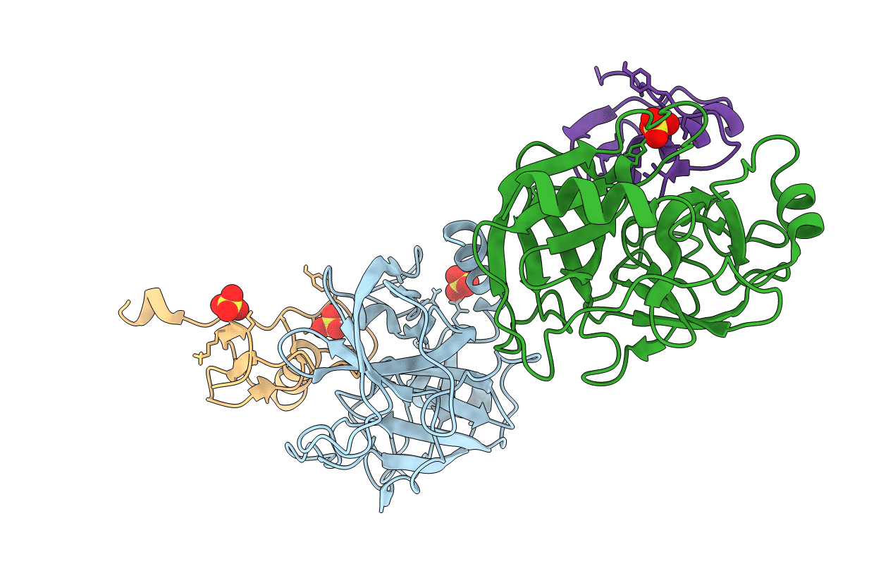
PDB ID:
7QE9
Keywords:
Title:
Human cationic trypsin (TRY1) complexed with serine protease inhibitor Kazal type 1 N34S (SPINK1 N34S)
Biological Source:
Source Organism(s):
Homo sapiens (Taxon ID: 9606)
Expression System(s):
Method Details:
Experimental Method:
Resolution:
2.10 Å
R-Value Free:
0.23
R-Value Work:
0.19
Space Group:
P 31 2 1
