7QAK image
Entry Detail
PDB ID:
7QAK
Keywords:
Title:
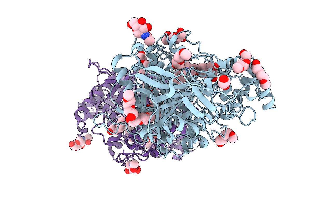
Mus Musculus Acetylcholinesterase in complex with 7-[(4-{[benzyl(methyl)amino]methyl}benzyl)oxy]-4-(hydroxymethyl)-2H-chromen-2-one
Biological Source:
Source Organism:
Host Organism:
PDB Version:
Deposition Date:
2021-11-17
Release Date:
2022-04-06
Method Details:
Experimental Method:
Resolution:
2.60 Å
R-Value Free:
0.22
R-Value Work:
0.18
R-Value Observed:
0.19
Space Group:
P 21 21 21
Macromolecular Entities
Polymer Type:polypeptide(L)
Description:Acetylcholinesterase
Chain IDs:A, B
Chain Length:543
Number of Molecules:2
Biological Source:Mus musculus
Primary Citation
Dual Reversible Coumarin Inhibitors Mutually Bound to Monoamine Oxidase B and Acetylcholinesterase Crystal Structures.
Acs Med.Chem.Lett. 13 499 506 (2022)
PMID: 35300078 DOI: 10.1021/acsmedchemlett.2c00001

Abstact

Multitarget directed ligands (MTDLs) represent a promising frontier in tackling the complexity of multifactorial pathologies. The synergistic inhibition of monoamine oxidase B (MAO B) and acetylcholinesterase (AChE) is believed to provide a potentiated effect in the treatment of Alzheimer's disease. Among previously reported micromolar or sub-micromolar coumarin-bearing dual inhibitors, compound 1 returned a tight-binding inhibition of MAO B (K i = 4.5 μM) and a +5.5 °C increase in the enzyme T m value. Indeed, the X-ray crystal structure revealed that binding of 1 produces unforeseen conformational changes at the MAO B entrance cavity. Interestingly, 1 showed great shape complementarity with the AChE enzymatic gorge, being deeply buried from the catalytic anionic subsite (CAS) to the peripheral anionic subsite (PAS) and causing significant structural changes in the active site. These findings provide structural templates for further development of dual MAO B and AChE inhibitors.

Legend

Protein

Chemical

Disease

Primary Citation of related structures