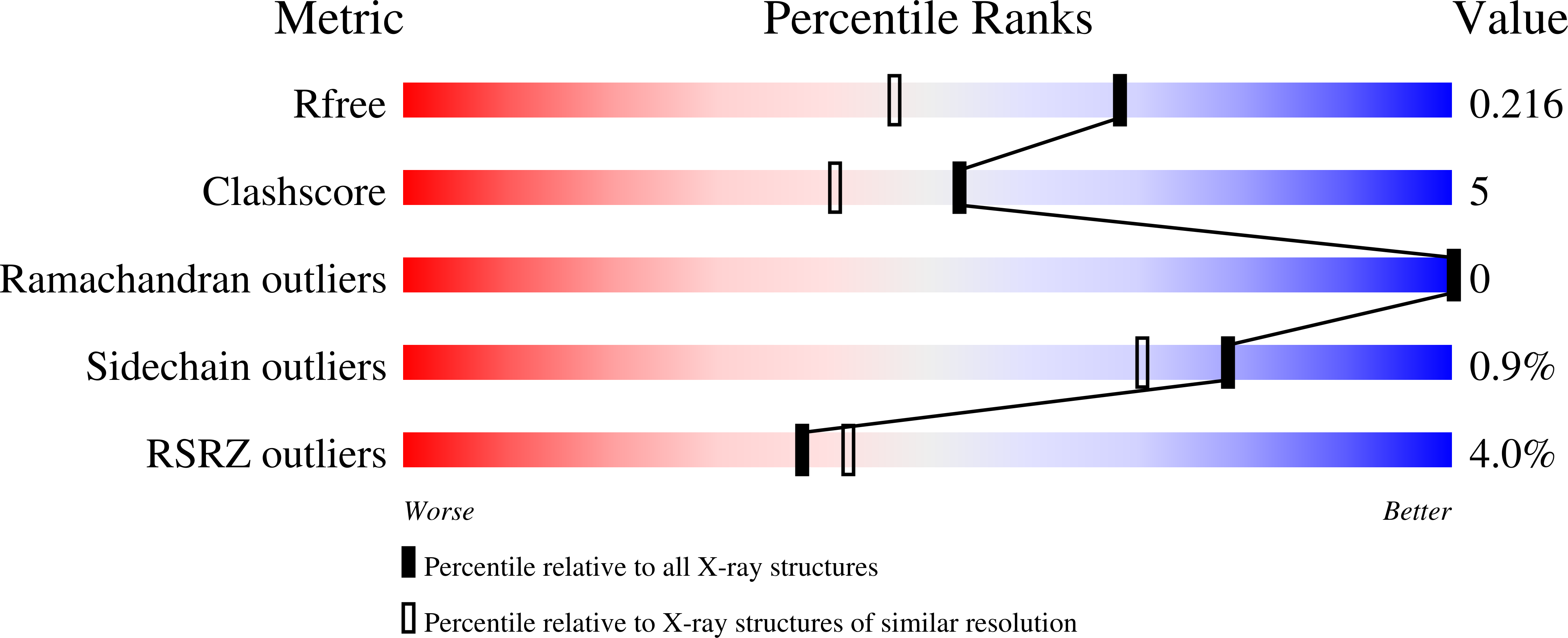
7Q9E image
Deposition Date 2021-11-12
Release Date 2022-10-19
Last Version Date 2024-01-31
Entry Detail
PDB ID:
7Q9E
Title:
CYP106A1
Biological Source:
Source Organism:
Host Organism:
Method Details:
Experimental Method:
Resolution:
1.70 Å
R-Value Free:
0.20
R-Value Work:
0.16
R-Value Observed:
0.17
Space Group:
P 1
Macromolecular Entities
Structures with similar UniProt ID
Protein Blast
Polymer Type:polypeptide(L)
Molecule:Cytochrome P450
Gene (Uniprot):BMD_1855
Chain IDs:A, B, C, D
Chain Length:416
Number of Molecules:4
Biological Source:Bacillus megaterium (strain DSM 319)
Primary Citation
Structural comparison of the cytochrome P450 enzymes CYP106A1 and CYP106A2 provides insight into their differences in steroid conversion.
Febs Lett. 596 3133 3144 (2022)
PMID: 36151590 DOI: 10.1002/1873-3468.14502

Abstact

Understanding the structural basis of the selectivity of steroid hydroxylation requires detailed structural and functional investigations on various steroid hydroxylases with different selectivities, such as the bacterial cytochrome P450 enzymes. Here, the crystal structure of the cytochrome P450 CYP106A1 from Priestia megaterium was solved. CYP106A1 exhibits a rare additional structural motif of a cytochrome P450, a sixth β-sheet. The protein was found in different unusual conformations corresponding to both open and closed forms even when crystallized without any known substrate. The structural comparison of CYP106A1 with the previously investigated CYP106A2, including docking studies for both isoforms with the substrate cortisol, reveals a completely different orientation of the steroid molecule in the active sites. This distinction convincingly explains the experimentally observed differences in substrate conversion and product formation by the two enzymes.

Legend

Protein

Chemical

Disease

Primary Citation of related structures