Deposition Date
2021-11-10
Release Date
2022-03-02
Last Version Date
2024-11-06
Entry Detail
PDB ID:
7Q84
Keywords:
Title:
Crystal structure of human peroxisomal acyl-Co-A oxidase 1a, apo-form
Biological Source:
Source Organism(s):
Homo sapiens (Taxon ID: 9606)
Expression System(s):
Method Details:
Experimental Method:
Resolution:
2.00 Å
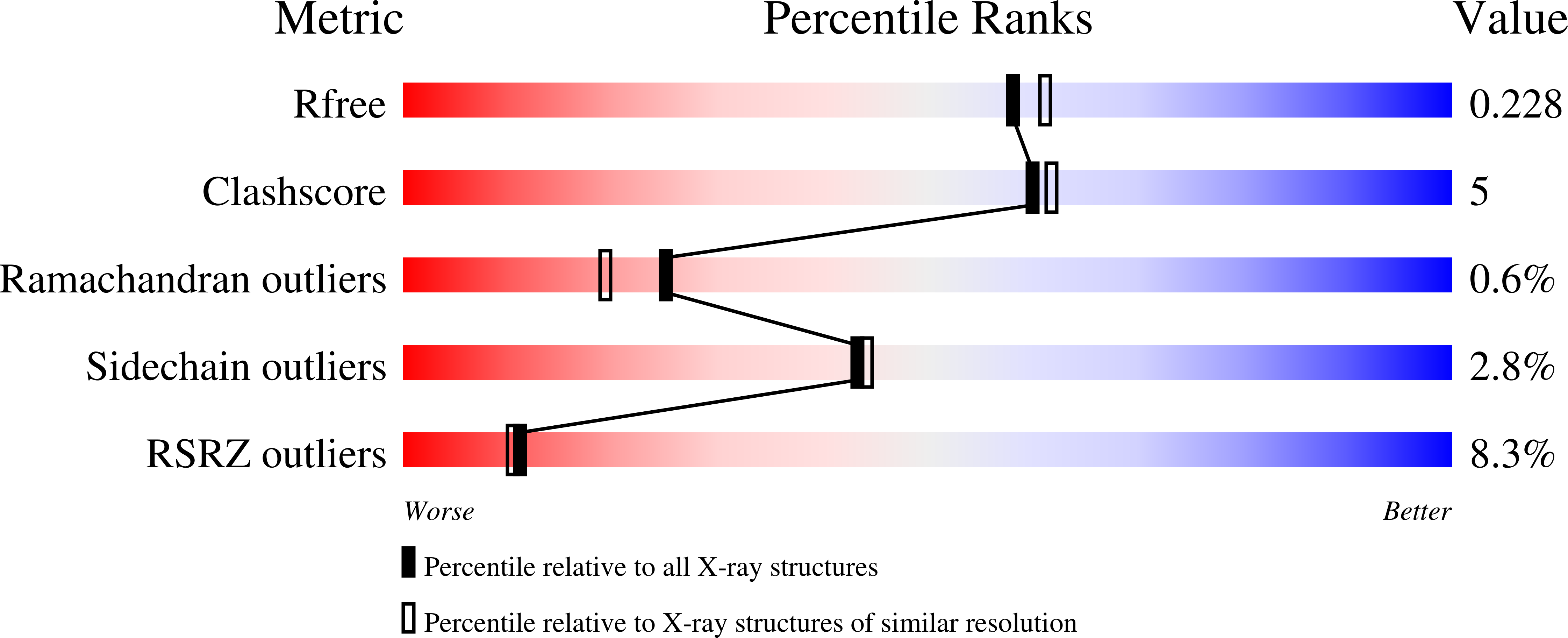
R-Value Free:
0.22
R-Value Work:
0.18
R-Value Observed:
0.18
Space Group:
P 21 21 21