7Q52 image
Deposition Date 2021-11-02
Release Date 2022-06-22
Last Version Date 2024-01-31
Entry Detail
PDB ID:
7Q52
Keywords:
Title:
Crystal structure of S/T protein kinase PknG from Mycobacterium tuberculosis in complex with inhibitor L2W
Biological Source:
Method Details:
Experimental Method:
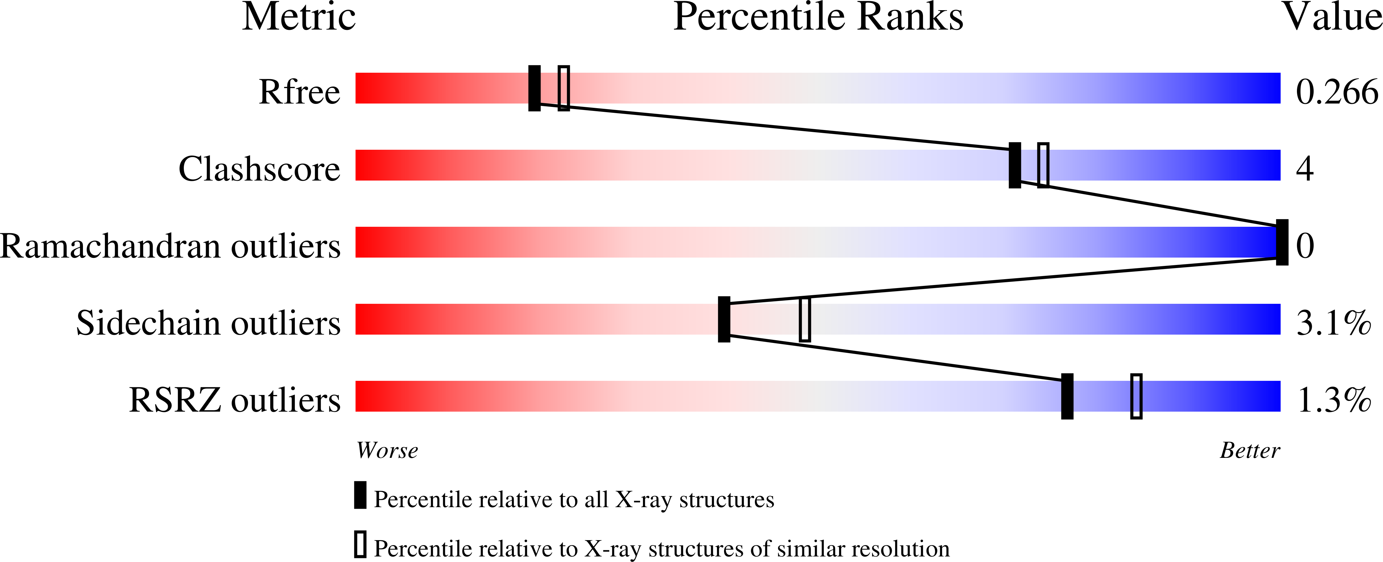
Resolution:
2.35 Å
R-Value Free:
0.26
R-Value Work:
0.21
Space Group:
C 1 2 1
Macromolecular Entities
Polymer Type:polypeptide(L)
Molecule:Serine/threonine-protein kinase PknG
Gene (Uniprot):pknG
Chain IDs:A (auth: AAA)
Chain Length:333
Number of Molecules:1
Biological Source:Mycobacterium tuberculosis (strain ATCC 25618 / H37Rv)
Primary Citation
Cosolvent Sites-Based Discovery of Mycobacterium Tuberculosis Protein Kinase G Inhibitors.
J.Med.Chem. 65 9691 9705 (2022)
PMID: 35737472 DOI: 10.1021/acs.jmedchem.1c02012

Abstact

Computer-aided drug discovery methods play a major role in the development of therapeutically important small molecules, but their performance needs to be improved. Molecular dynamics simulations in mixed solvents are useful in understanding protein-ligand recognition and improving molecular docking predictions. In this work, we used ethanol as a cosolvent to find relevant interactions for ligands toward protein kinase G, an essential protein of Mycobacterium tuberculosis (Mtb). We validated the hot spots by screening a database of fragment-like compounds and another one of known kinase inhibitors. Next, we performed a pharmacophore-guided docking simulation and found three low micromolar inhibitors, including one with a novel chemical scaffold that we expanded to four derivative compounds. Binding affinities were characterized by intrinsic fluorescence quenching assays, isothermal titration calorimetry, and the analysis of melting curves. The predicted binding mode was confirmed by X-ray crystallography. Finally, the compounds significantly inhibited the viability of Mtb in infected THP-1 macrophages.

Legend

Protein

Chemical

Disease

Primary Citation of related structures