7PZH image
Deposition Date 2021-10-12
Release Date 2022-05-25
Last Version Date 2024-01-31
Entry Detail
PDB ID:
7PZH
Keywords:
Title:
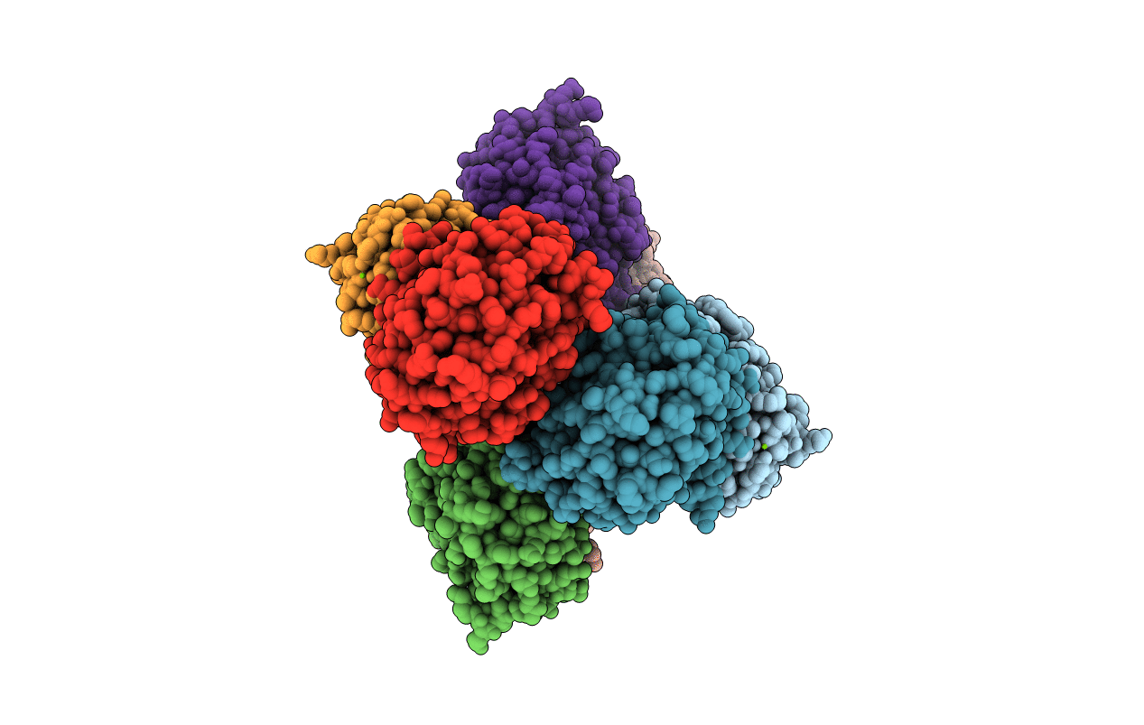
Phocaeicola vulgatus sialic acid esterase at 2.06 Angstrom resolution
Biological Source:
Source Organism:
Method Details:
Experimental Method:
Resolution:
2.06 Å
R-Value Free:
0.22
R-Value Work:
0.18
Space Group:
P 61
Macromolecular Entities
Structures with similar UniProt ID
Protein Blast
Polymer Type:polypeptide(L)
Molecule:Lysophospholipase L1
Gene (Uniprot):DWX04_04065, ERS852457_01148, F9Z94_13025, GAS37_03660, GAY17_10630, GAY79_04625, GAY98_11740, GAZ06_07335, GAZ09_07170, GAZ76_12675, GAZ92_18925, HKQ51_19175, HKQ55_14115, HUV05_02570, RVY68_15925
Chain IDs:A (auth: AAA), B (auth: BBB), C (auth: CCC), D (auth: DDD), E (auth: EEE), F (auth: FFF), G (auth: HHH), H (auth: GGG)
Chain Length:221
Number of Molecules:8
Biological Source:Phocaeicola vulgatus
Ligand Molecules
Primary Citation
The structure of Phocaeicola vulgatus sialic acid acetylesterase.
Acta Crystallogr D Struct Biol 78 647 657 (2022)
PMID: 35503212 DOI: 10.1107/S2059798322003357

Abstact

Sialic acids terminate many N- and O-glycans and are widely distributed on cell surfaces. There are a diverse range of enzymes which interact with these sugars throughout the tree of life. They can act as receptors for influenza and specific betacoronaviruses in viral binding and their cleavage is important in virion release. Sialic acids are also exploited by both commensal and pathogenic bacteria for nutrient acquisition. A common modification of sialic acid is 9-O-acetylation, which can limit the action of sialidases. Some bacteria, including human endosymbionts, employ esterases to overcome this modification. However, few bacterial sialic acid 9-O-acetylesterases (9-O-SAEs) have been structurally characterized. Here, the crystal structure of a 9-O-SAE from Phocaeicola vulgatus (PvSAE) is reported. The structure of PvSAE was determined to resolutions of 1.44 and 2.06 Å using crystals from two different crystallization conditions. Structural characterization revealed PvSAE to be a dimer with an SGNH fold, named after the conserved sequence motif of this family, and a Ser-His-Asp catalytic triad. These structures also reveal flexibility in the most N-terminal α-helix, which provides a barrier to active-site accessibility. Biochemical assays also show that PvSAE deacetylates both mucin and the acetylated chromophore para-nitrophenyl acetate. This structural and biochemical characterization of PvSAE furthers the understanding of 9-O-SAEs and may aid in the discovery of small molecules targeting this class of enzyme.

Legend

Protein

Chemical

Disease

Primary Citation of related structures
Feedback Form
Name
Email
Institute
Feedback