7PV9 image
Entry Detail
PDB ID:
7PV9
Keywords:
Title:
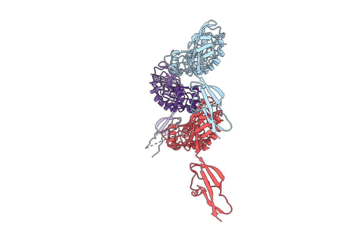
Listeria monocytogene InlB (internalin B) residues 36-392 (internalin domain and B-repeat)
Biological Source:
PDB Version:
Deposition Date:
2021-10-01
Release Date:
2022-01-26
Method Details:
Experimental Method:
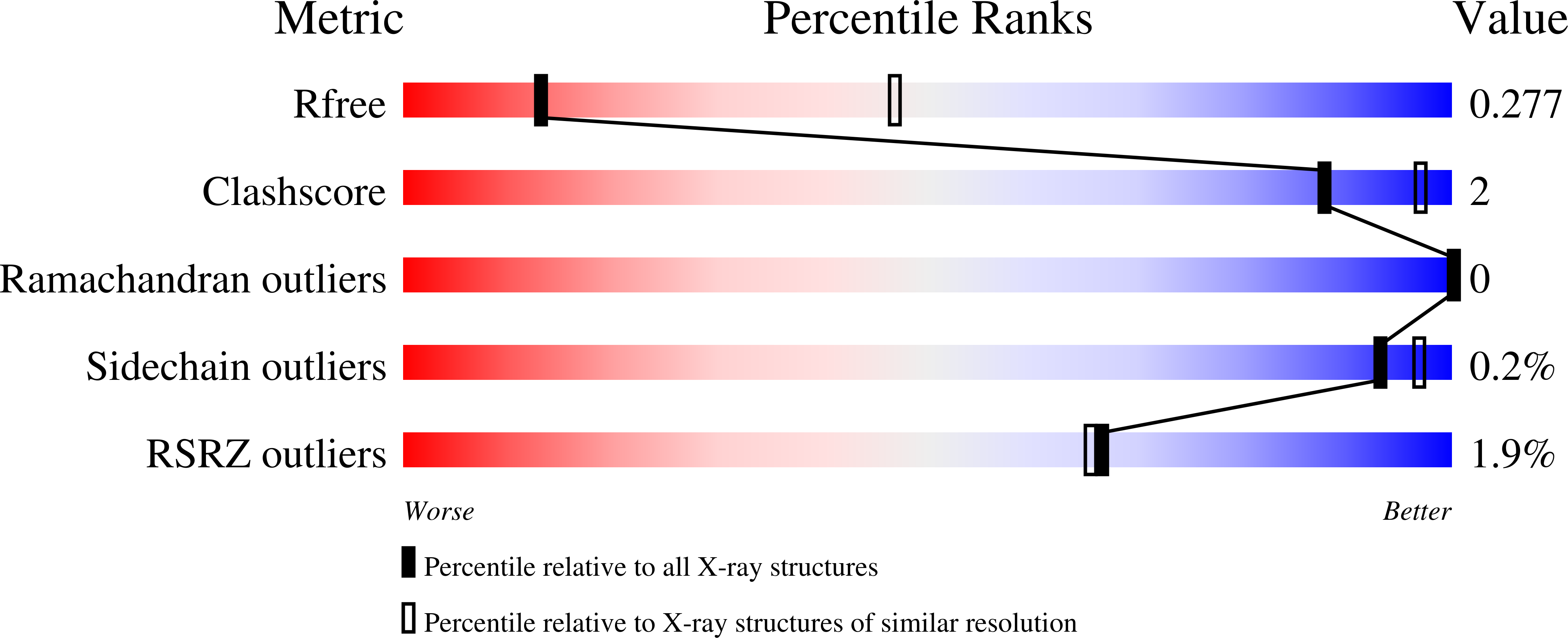
Resolution:
3.30 Å
R-Value Free:
0.27
R-Value Work:
0.23
R-Value Observed:
0.23
Space Group:
P 21 21 21
Macromolecular Entities
Polymer Type:polypeptide(L)
Description:Internalin B
Chain IDs:A, B, C
Chain Length:362
Number of Molecules:3
Biological Source:Listeria monocytogenes serovar 1/2a (strain ATCC BAA-679 / EGD-e)
Ligand Molecules
Primary Citation
A recurring packing contact in crystals of InlB pinpoints functional binding sites in the internalin domain and the B repeat.
Acta Crystallogr D Struct Biol 78 310 320 (2022)
PMID: 35234145 DOI: 10.1107/S2059798322000432

Abstact

InlB, a bacterial agonist of the human receptor tyrosine kinase MET, consists of an N-terminal internalin domain, a central B repeat and three C-terminal GW domains. In all previous structures of full-length InlB or an InlB construct lacking the GW domains (InlB392), there was no interpretable electron density for the B repeat. Here, three InlB392 crystal structures in which the B repeat is resolved are described. These are the first structures to reveal the relative orientation of the internalin domain and the B repeat. A wild-type structure and two structures of the T332E variant together contain five crystallographically independent molecules. Surprisingly, the threonine-to-glutamate substitution in the B repeat substantially improved the crystallization propensity and crystal quality of the T332E variant. The internalin domain and B repeat are quite rigid internally, but are flexibly linked to each other. The new structures show that inter-domain flexibility is the most likely cause of the missing electron density for the B repeat in previous InlB structures. A potential binding groove between B-repeat strand β2 and an adjacent loop forms an important crystal contact in all five crystallographically independent chains. This region may represent a hydrophobic `sticky patch' that supports protein-protein interactions. This assumption agrees with the previous finding that all known inactivating point mutations in the B repeat lie within strand β2. The groove formed by strand β2 and the adjacent loop may thus represent a functionally important protein-protein interaction site in the B repeat.

Legend

Protein

Chemical

Disease

Primary Citation of related structures