Deposition Date
2021-09-29
Release Date
2022-01-19
Last Version Date
2024-01-31
Entry Detail
Biological Source:
Source Organism(s):
uncultured bacterium (Taxon ID: 77133)
Expression System(s):
Method Details:
Experimental Method:
Resolution:
2.66 Å
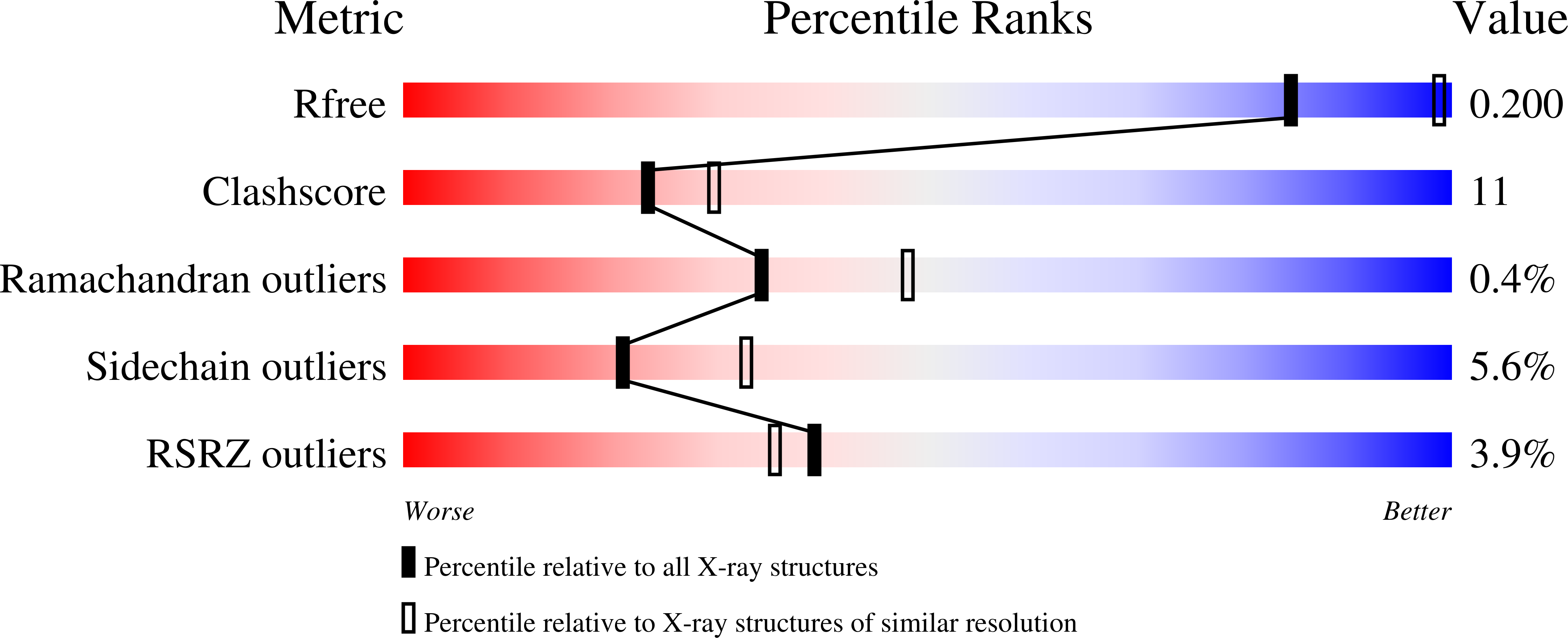
R-Value Free:
0.21
R-Value Work:
0.17
R-Value Observed:
0.17
Space Group:
P 61 2 2