7PSY image
Deposition Date 2021-09-24
Release Date 2022-01-12
Last Version Date 2024-01-31
Entry Detail
PDB ID:
7PSY
Title:
X-ray crystal structure of perdeuterated LecB lectin in complex with perdeuterated fucose
Biological Source:
Source Organism:
Method Details:
Experimental Method:
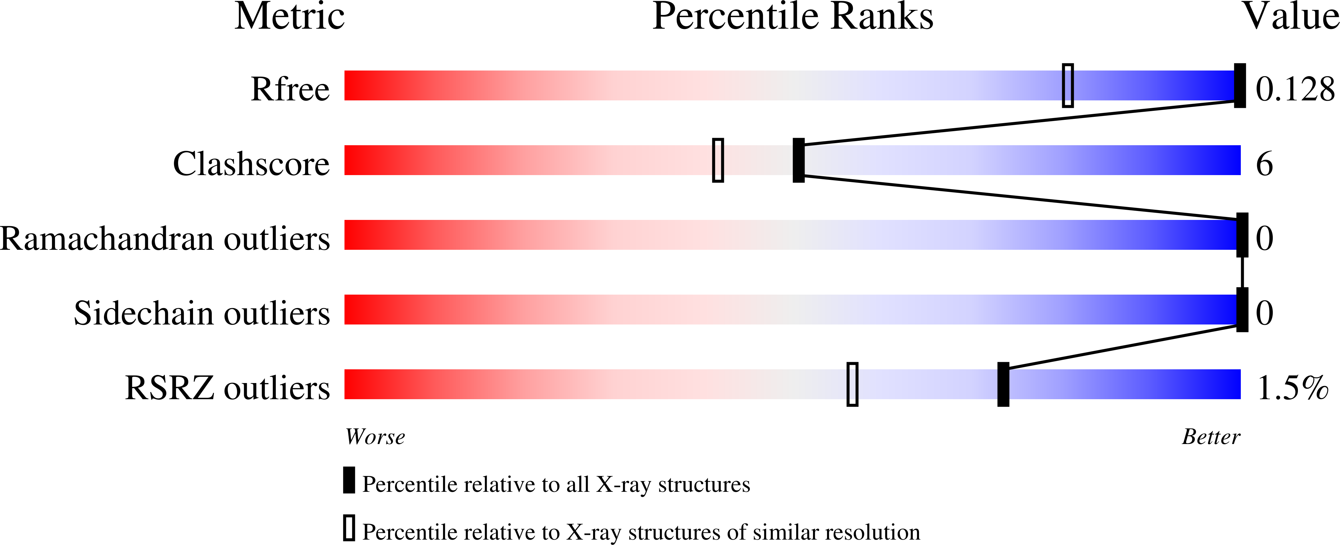
Resolution:
0.90 Å
R-Value Free:
0.13
R-Value Work:
0.11
R-Value Observed:
0.11
Space Group:
P 1 21 1
Macromolecular Entities
Structures with similar UniProt ID
Protein Blast
Polymer Type:polypeptide(L)
Molecule:Fucose-binding lectin
Chain IDs:A, B, C, D
Chain Length:115
Number of Molecules:4
Biological Source:Pseudomonas aeruginosa
Primary Citation
Neutron crystallography reveals mechanisms used by Pseudomonas aeruginosa for host-cell binding.
Nat Commun 13 194 194 (2022)
PMID: 35017516 DOI: 10.1038/s41467-021-27871-8

Abstact

The opportunistic pathogen Pseudomonas aeruginosa, a major cause of nosocomial infections, uses carbohydrate-binding proteins (lectins) as part of its binding to host cells. The fucose-binding lectin, LecB, displays a unique carbohydrate-binding site that incorporates two closely located calcium ions bridging between the ligand and protein, providing specificity and unusually high affinity. Here, we investigate the mechanisms involved in binding based on neutron crystallography studies of a fully deuterated LecB/fucose/calcium complex. The neutron structure, which includes the positions of all the hydrogen atoms, reveals that the high affinity of binding may be related to the occurrence of a low-barrier hydrogen bond induced by the proximity of the two calcium ions, the presence of coordination rings between the sugar, calcium and LecB, and the dynamic behaviour of bridging water molecules at room temperature. These key structural details may assist in the design of anti-adhesive compounds to combat multi-resistance bacterial infections.

Legend

Protein

Chemical

Disease

Primary Citation of related structures