
Deposition Date
2021-09-24
Release Date
2022-07-20
Last Version Date
2024-01-31
Entry Detail
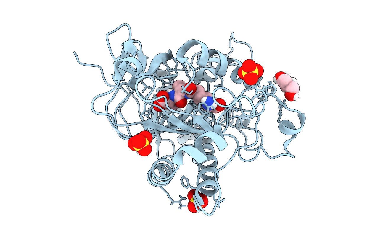
PDB ID:
7PSW
Keywords:
Title:
Spin labeled IPNS S55C variant in complex with Fe and ACV under anaerobic conditions
Biological Source:
Source Organism(s):
Expression System(s):
Method Details:
Experimental Method:
Resolution:
1.21 Å
R-Value Free:
0.16
R-Value Work:
0.14
R-Value Observed:
0.14
Space Group:
P 21 21 21
