7PRC image
Entry Detail
PDB ID:
7PRC
Title:
PHOTOSYNTHETIC REACTION CENTER FROM RHODOPSEUDOMONAS VIRIDIS (DG-420315 (TRIAZINE) COMPLEX)
Biological Source:
Source Organism:
PDB Version:
Deposition Date:
1997-08-01
Release Date:
1999-04-06
Method Details:
Experimental Method:
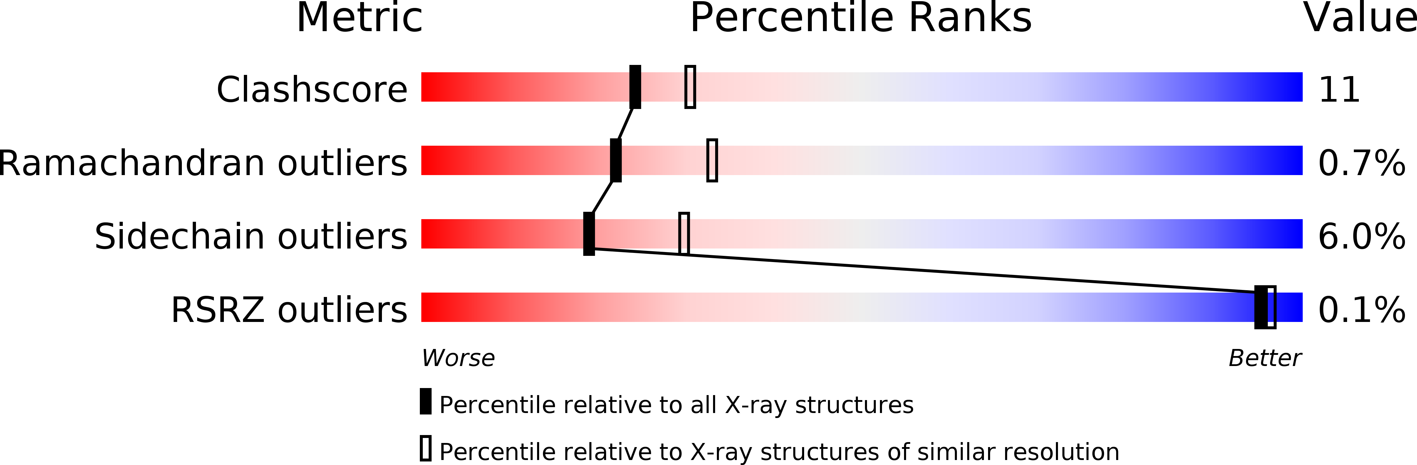
Resolution:
2.65 Å
R-Value Free:
0.23
R-Value Work:
0.18
R-Value Observed:
0.18
Space Group:
P 43 21 2
Macromolecular Entities
Polymer Type:polypeptide(L)
Description:PHOTOSYNTHETIC REACTION CENTER
Chain IDs:A (auth: C)
Chain Length:336
Number of Molecules:1
Biological Source:Blastochloris viridis
Polymer Type:polypeptide(L)
Description:PHOTOSYNTHETIC REACTION CENTER
Chain IDs:D (auth: H)
Chain Length:258
Number of Molecules:1
Biological Source:Blastochloris viridis
Polymer Type:polypeptide(L)
Description:PHOTOSYNTHETIC REACTION CENTER
Chain IDs:B (auth: L)
Chain Length:273
Number of Molecules:1
Biological Source:Blastochloris viridis
Polymer Type:polypeptide(L)
Description:PHOTOSYNTHETIC REACTION CENTER
Chain IDs:C (auth: M)
Chain Length:323
Number of Molecules:1
Biological Source:Blastochloris viridis
Modified Residue
Compound ID Chain ID Parent Comp ID Details 2D Image
FME D MET N-FORMYLMETHIONINE
Primary Citation
Refined crystal structures of reaction centres from Rhodopseudomonas viridis in complexes with the herbicide atrazine and two chiral atrazine derivatives also lead to a new model of the bound carotenoid.
J.Mol.Biol. 286 883 898 (1999)
PMID: 10024457 DOI: 10.1006/jmbi.1998.2532

Abstact

In a reaction of central importance to the energetics of photosynthetic bacteria, light-induced electron transfer in the reaction centre (RC) is coupled with the uptake of protons from the cytoplasm at the binding site of the secondary quinone (QB). It has been established by X-ray crystallography that the triazine herbicide terbutryn binds to the QB site. However, the exact description of protein-triazine interactions has had to await the refinement of higher-resolution structures. In addition, there is also interest in the role of chirality in the activity of herbicides. Here, we report the structural characterisation of triazine binding by crystallographic refinement of complexes of the RC either with the triazine inhibitor atrazine (Protein Data Bank (PDB) entry 5PRC) or with the chiral atrazine derivatives, DG-420314 (S(-) enantiomer, PDB entry 6PRC) or DG-420315 (R(+) enantiomer, PDB entry 7PRC). Due to the high quality of the data collected, it has been possible to describe the exact nature of triazine binding and its effect on the structure of the protein at high-resolution limits of 2.35 A (5PRC), 2.30 A (6PRC), and 2.65 A (7PRC), respectively. In addition to two previously implied hydrogen bonds, a third hydrogen bond, binding the distal side of the inhibitors to the protein, and four additional hydrogen bonds mediated by two tightly bound water molecules on the proximal side of the inhibitors, are apparent. Based on the high quality data collected on the RC complexes of the two chiral atrazine derivatives, unequivocal assignment of the structure at the chiral centres was possible, even though the differences in structures of the substituents are small. The structures provide explanations for the relative binding affinities of the two chiral compounds. Although it was not an explicit goal of this work, the new data were of sufficient quality to improve the original model also regarding the structure of the bound carotenoid 1,2-dihydroneurosporene. A carotenoid model with a cis double bond at the 15,15' position fits the electron density better than the original model with a 13,14-cis double bond.

Legend

Protein

Chemical

Disease

Primary Citation of related structures