7PP3 image
Entry Detail
PDB ID:
7PP3
Keywords:
Title:
STRUCTURE OF ESTER-HYDROLASE EH7 FROM THE METAGENOME OF MARINE SEDIMENTS AT MILAZZO HARBOR (SICILY, ITALY)
Biological Source:
Source Organism:
PDB Version:
Deposition Date:
2021-09-13
Release Date:
2022-07-06
Method Details:
Experimental Method:
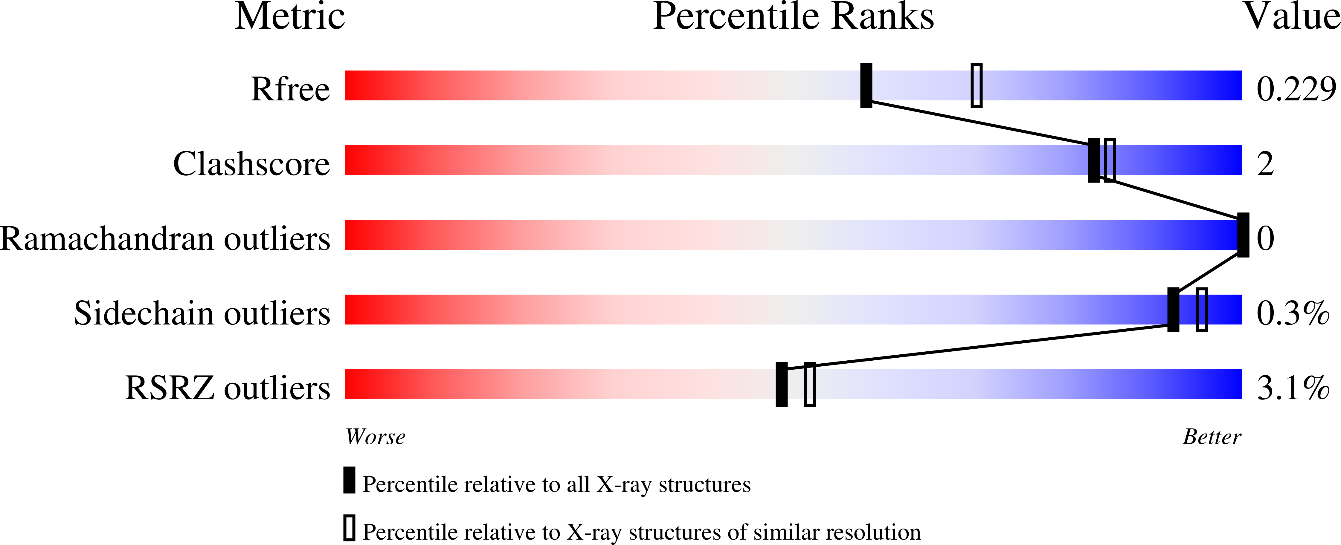
Resolution:
2.25 Å
R-Value Free:
0.22
R-Value Work:
0.18
R-Value Observed:
0.18
Space Group:
P 43 2 2
Macromolecular Entities
Polymer Type:polypeptide(L)
Description:Esterase
Chain IDs:A, B, C, D
Chain Length:429
Number of Molecules:4
Biological Source:uncultured bacterium
Primary Citation
Crystal structure of a family VIII beta-lactamase fold hydrolase reveals the molecular mechanism for its broad substrate scope.
Febs J. 289 6714 6730 (2022)
PMID: 35694902 DOI: 10.1111/febs.16554

Abstact

Family VIII esterases present similarities to class C β-lactamases, which show nucleophilic serines located at the S-X-X-K motif instead of the G-X-S-X-G or G-D-S-(L) motif shown by other carboxylesterase families. Here, we report the crystal structure of a novel family VIII (subfamily VIII. I) esterase (EH7 ; denaturing temperature, 52.6 ± 0.3 °C; pH optimum 7.0-9.0) to deepen its broad substrate range. Indeed, the analysis of the substrate specificity revealed its capacity to hydrolyse nitrocefin as a model chromogenic cephalosporin substrate (40.4 ± 11.4 units·g-1), and a large battery of 66 structurally different esters (up to 1730 min-1), including bis(2-hydroxyethyl)-terephthalate (241.7 ± 8.5 units·g-1) and the mycotoxin T-2 (1220 ± 52 units·g-1). It also showed acyltransferase activity through the synthesis of benzyl 3-oxobutanoate (40.4 ± 11.4 units·g-1) from benzyl alcohol and vinyl acetoacetate. Such a broad substrate scope is rare among family VIII esterases and lipolytic enzymes. Structural analyses of free and substrate-bound forms of this homooctamer esterase suggest that EH7 presents a more opened and exposed S1 site having no steric hindrance for the entrance of substrates to the active site, more flexible R1, R2 and R3 regions allowing for the binding of a wide spectrum of substrates into the active site, and small residues in the conserved motif Y-X-X containing the catalytic Tyr enabling the entrance of large substrates. These unique structural elements in combination with docking experiments allowed us to gain valuable insights into the substrate specificity of this esterase and possible others belonging to family VIII.

Legend

Protein

Chemical

Disease

Primary Citation of related structures