Deposition Date
2021-08-23
Release Date
2021-12-22
Last Version Date
2024-10-16
Entry Detail
PDB ID:
7PJD
Keywords:
Title:
The X-ray structure of juvenile hormone diol kinase from the silk worm Bombyx mori.
Biological Source:
Source Organism(s):
Bombyx mori (Taxon ID: 7091)
Expression System(s):
Method Details:
Experimental Method:
Resolution:
1.99 Å
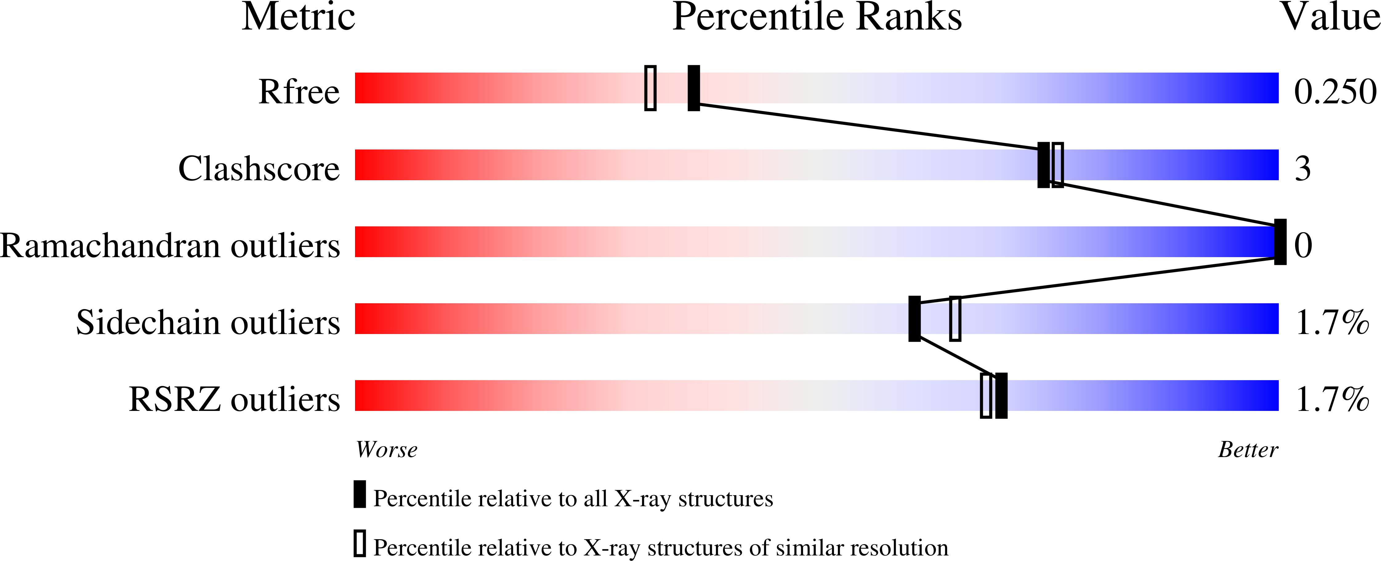
R-Value Free:
0.24
R-Value Work:
0.19
R-Value Observed:
0.19
Space Group:
P 1 21 1