7PJ6 image
Deposition Date 2021-08-23
Release Date 2022-09-07
Last Version Date 2024-02-07
Entry Detail
PDB ID:
7PJ6
Keywords:
Title:
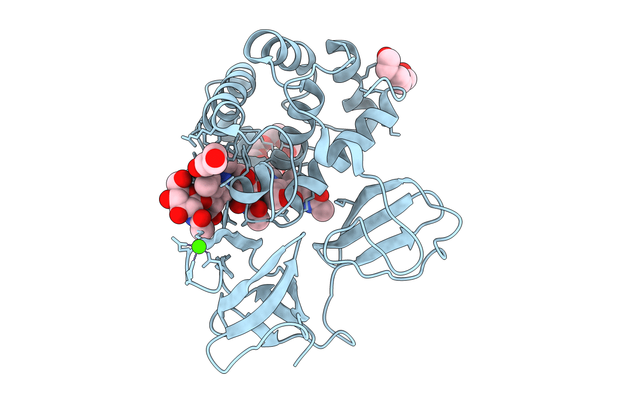
Crystal structure of catalytic domain of LytB (E585Q) from Streptococcus pneumoniae in complex with NAG-NAM-NAG-NAM-NAG peptidolycan analogue
Biological Source:
Method Details:
Experimental Method:
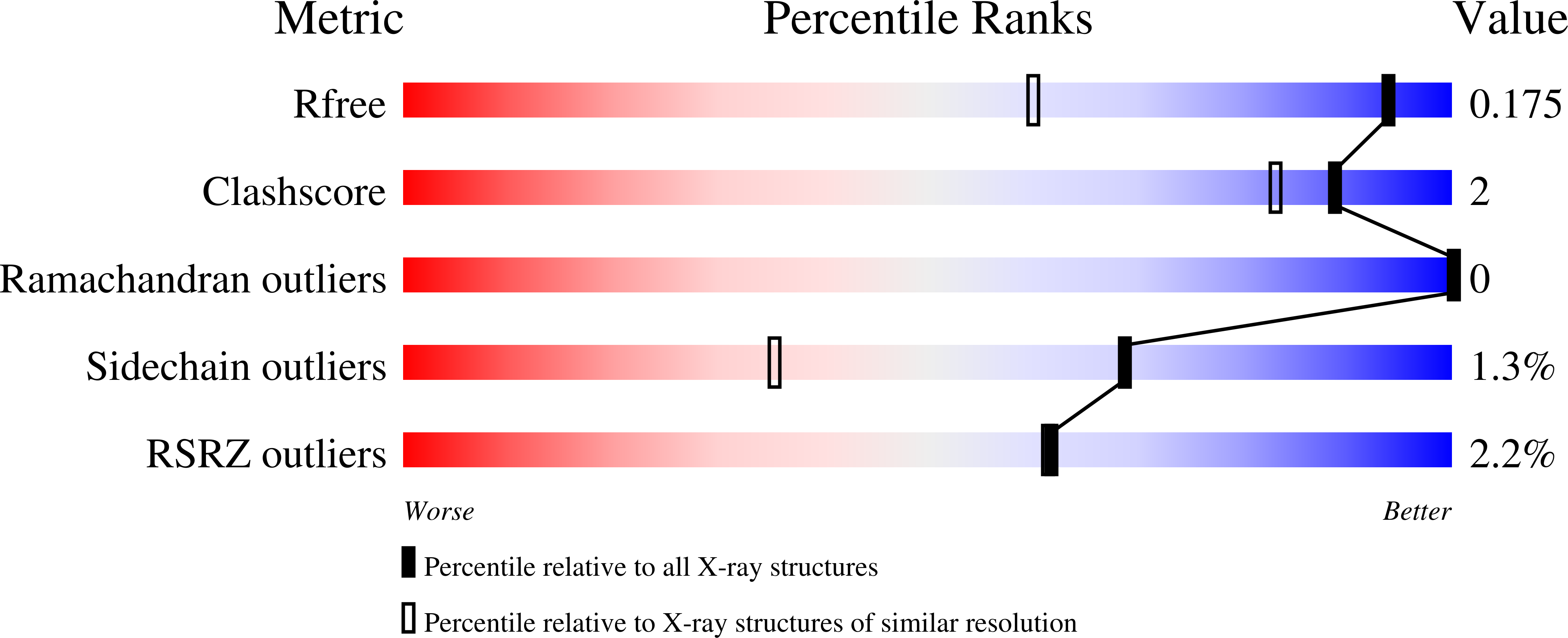
Resolution:
1.30 Å
R-Value Free:
0.17
R-Value Work:
0.14
Space Group:
C 2 2 21
Macromolecular Entities
Polymer Type:polypeptide(L)
Molecule:Putative endo-beta-N-acetylglucosaminidase
Gene (Uniprot):lytB
Mutations:E585Q
Chain IDs:A (auth: AAA)
Chain Length:274
Number of Molecules:1
Biological Source:Streptococcus pneumoniae (strain ATCC BAA-255 / R6)
Primary Citation
Molecular basis of the final step of cell division in Streptococcus pneumoniae.
Cell Rep 42 112756 112756 (2023)
PMID: 37418323 DOI: 10.1016/j.celrep.2023.112756

Abstact

Bacterial cell-wall hydrolases must be tightly regulated during bacterial cell division to prevent aberrant cell lysis and to allow final separation of viable daughter cells. In a multidisciplinary work, we disclose the molecular dialogue between the cell-wall hydrolase LytB, wall teichoic acids, and the eukaryotic-like protein kinase StkP in Streptococcus pneumoniae. After characterizing the peptidoglycan recognition mode by the catalytic domain of LytB, we further demonstrate that LytB possesses a modular organization allowing the specific binding to wall teichoic acids and to the protein kinase StkP. Structural and cellular studies notably reveal that the temporal and spatial localization of LytB is governed by the interaction between specific modules of LytB and the final PASTA domain of StkP. Our data collectively provide a comprehensive understanding of how LytB performs final separation of daughter cells and highlights the regulatory role of eukaryotic-like kinases on lytic machineries in the last step of cell division in streptococci.

Legend

Protein

Chemical

Disease

Primary Citation of related structures