Deposition Date
2021-08-09
Release Date
2021-10-13
Last Version Date
2024-11-06
Entry Detail
PDB ID:
7PEE
Keywords:
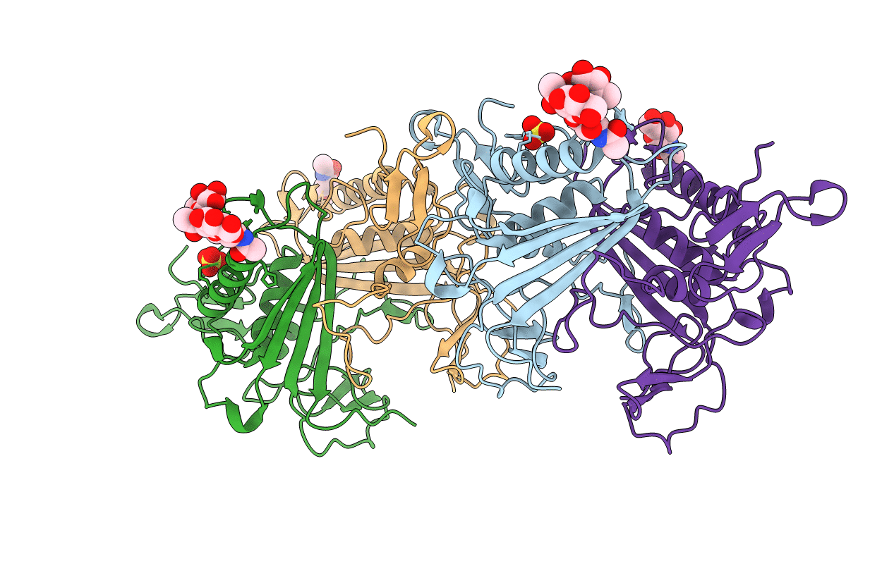
Title:
Crystal structure of extracellular part of human Trop2
Biological Source:
Source Organism(s):
Homo sapiens (Taxon ID: 9606)
Expression System(s):
Method Details:
Experimental Method:
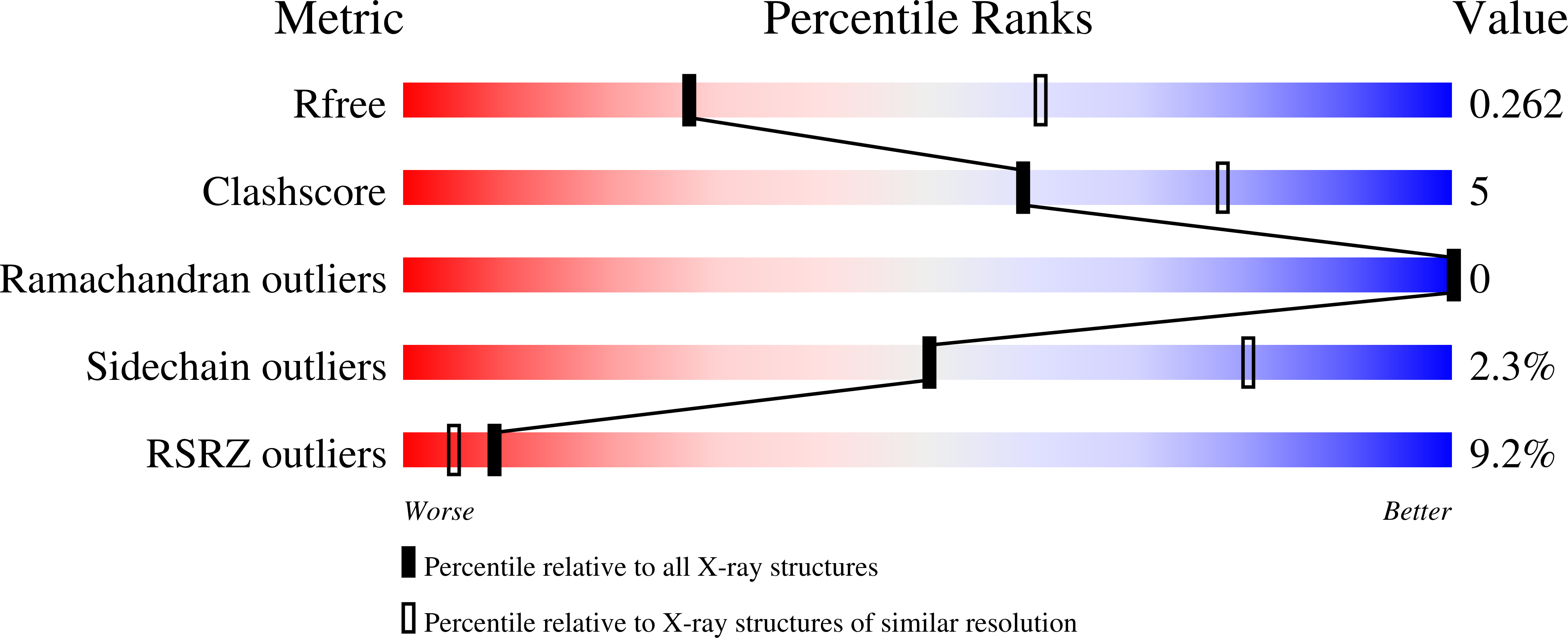
Resolution:
2.81 Å
R-Value Free:
0.26
R-Value Work:
0.23
R-Value Observed:
0.24
Space Group:
P 43 2 2