7PD2 image
Entry Detail
PDB ID:
7PD2
Keywords:
Title:
Crystal structure of the substrate-free radical SAM tyrosine lyase ThiH (2-iminoacetate synthase) from Thermosinus carboxydivorans
Biological Source:
PDB Version:
Deposition Date:
2021-08-04
Release Date:
2022-05-11
Method Details:
Experimental Method:
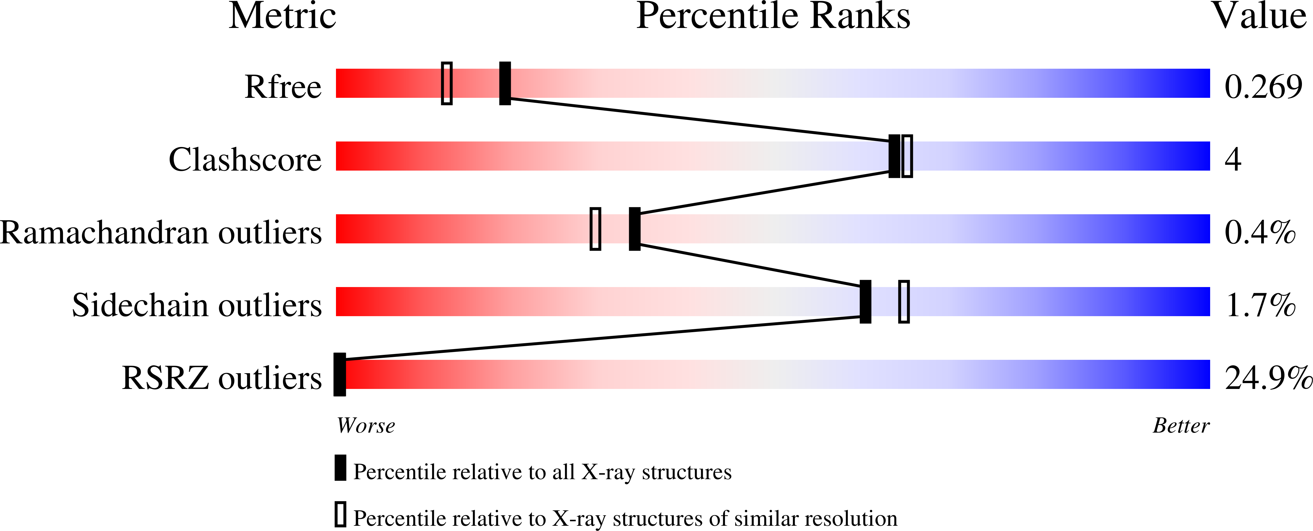
Resolution:
1.99 Å
R-Value Free:
0.26
R-Value Work:
0.22
R-Value Observed:
0.23
Space Group:
C 1 2 1
Macromolecular Entities
Polymer Type:polypeptide(L)
Description:Thiazole biosynthesis protein ThiH
Chain IDs:A, B
Chain Length:377
Number of Molecules:2
Biological Source:Thermosinus carboxydivorans Nor1
Primary Citation
L-tyrosine-bound ThiH structure reveals C-C bond break differences within radical SAM aromatic amino acid lyases.
Nat Commun 13 2284 2284 (2022)
PMID: 35477710 DOI: 10.1038/s41467-022-29980-4

Abstact

2-iminoacetate synthase ThiH is a radical S-adenosyl-L-methionine (SAM) L-tyrosine lyase and catalyzes the L-tyrosine Cα-Cβ bond break to produce dehydroglycine and p-cresol while the radical SAM L-tryptophan lyase NosL cleaves the L-tryptophan Cα-C bond to produce 3-methylindole-2-carboxylic acid. It has been difficult to understand the features that condition one C-C bond break over the other one because the two enzymes display significant primary structure similarities and presumably similar substrate-binding modes. Here, we report the crystal structure of L-tyrosine bound ThiH from Thermosinus carboxydivorans revealing an unusual protonation state of L-tyrosine upon binding. Structural comparison of ThiH with NosL and computational studies of the respective reactions they catalyze show that substrate activation is eased by tunneling effect and that subtle structural changes between the two enzymes affect, in particular, the hydrogen-atom abstraction by the 5´-deoxyadenosyl radical species, driving the difference in reaction specificity.

Legend

Protein

Chemical

Disease

Primary Citation of related structures