7PBW image
Deposition Date 2021-08-02
Release Date 2021-11-24
Last Version Date 2024-11-20
Entry Detail
PDB ID:
7PBW
Keywords:
Title:
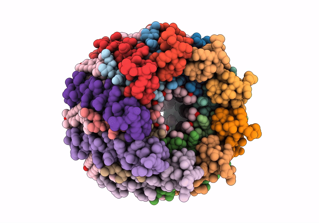
Cryo-EM structure of light harvesting complex 2 from Rba. sphaeroides.
Biological Source:
Source Organism:
Method Details:
Experimental Method:
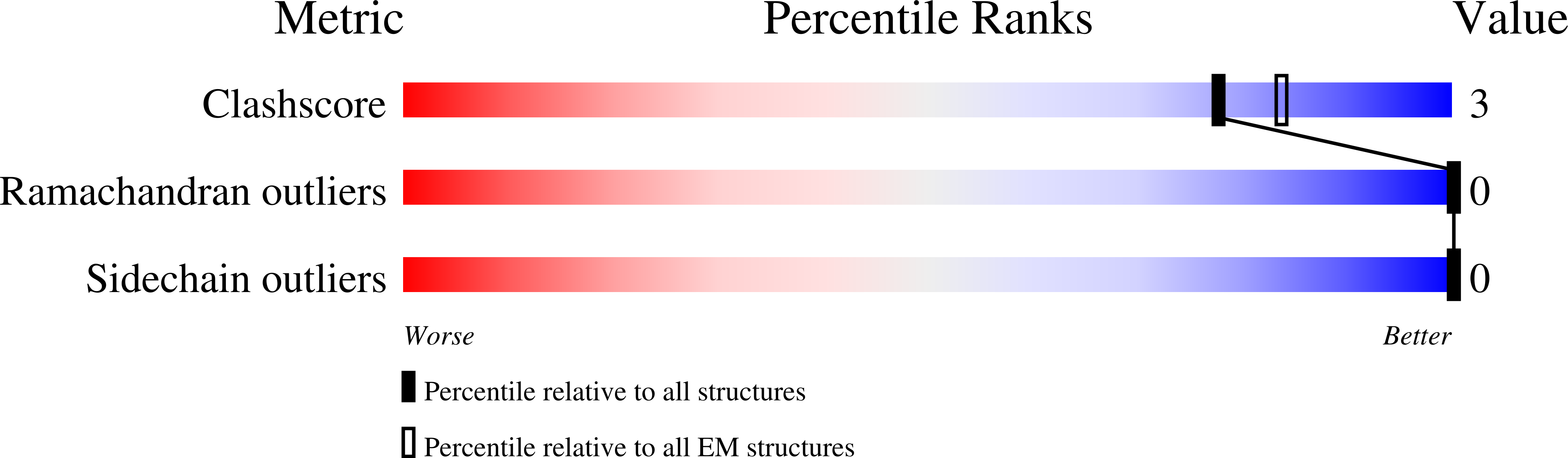
Resolution:
2.10 Å
Aggregation State:
PARTICLE
Reconstruction Method:
SINGLE PARTICLE
Macromolecular Entities
Structures with similar UniProt ID
Protein Blast
Polymer Type:polypeptide(L)
Molecule:Light-harvesting protein B-800/850 alpha chain
Gene (Uniprot):pucA
Chain IDs:A (auth: AA), B (auth: AB), C (auth: AC), D (auth: AD), E (auth: AE), F (auth: AF), G (auth: AG), H (auth: AH), I (auth: AI)
Chain Length:50
Number of Molecules:9
Biological Source:Cereibacter sphaeroides 2.4.1
Structures with similar UniProt ID
Protein Blast
Polymer Type:polypeptide(L)
Molecule:Light-harvesting protein B-800/850 beta chain
Gene (Uniprot):pucB
Chain IDs:J (auth: BA), K (auth: BB), L (auth: BC), M (auth: BD), N (auth: BE), O (auth: BF), P (auth: BG), Q (auth: BH), R (auth: BI)
Chain Length:47
Number of Molecules:9
Biological Source:Cereibacter sphaeroides 2.4.1
Modified Residue
Compound ID Chain ID Parent Comp ID Details 2D Image
CXM A MET modified residue
Primary Citation
Cryo-EM Structure of the Rhodobacter sphaeroides Light-Harvesting 2 Complex at 2.1 angstrom.
Biochemistry 60 3302 3314 (2021)
PMID: 34699186 DOI: 10.1021/acs.biochem.1c00576

Abstact

Light-harvesting 2 (LH2) antenna complexes augment the collection of solar energy in many phototrophic bacteria. Despite its frequent role as a model for such complexes, there has been no three-dimensional (3D) structure available for the LH2 from the purple phototroph Rhodobacter sphaeroides. We used cryo-electron microscopy (cryo-EM) to determine the 2.1 Å resolution structure of this LH2 antenna, which is a cylindrical assembly of nine αβ heterodimer subunits, each of which binds three bacteriochlorophyll a (BChl) molecules and one carotenoid. The high resolution of this structure reveals all of the interpigment and pigment-protein interactions that promote the assembly and energy-transfer properties of this complex. Near the cytoplasmic face of the complex there is a ring of nine BChls, which absorb maximally at 800 nm and are designated as B800; each B800 is coordinated by the N-terminal carboxymethionine of LH2-α, part of a network of interactions with nearby residues on both LH2-α and LH2-β and with the carotenoid. Nine carotenoids, which are spheroidene in the strain we analyzed, snake through the complex, traversing the membrane and interacting with a ring of 18 BChls situated toward the periplasmic side of the complex. Hydrogen bonds with C-terminal aromatic residues modify the absorption of these pigments, which are red-shifted to 850 nm. Overlaps between the macrocycles of the B850 BChls ensure rapid transfer of excitation energy around this ring of pigments, which act as the donors of energy to neighboring LH2 and reaction center light-harvesting 1 (RC-LH1) complexes.

Legend

Protein

Chemical

Disease

Primary Citation of related structures