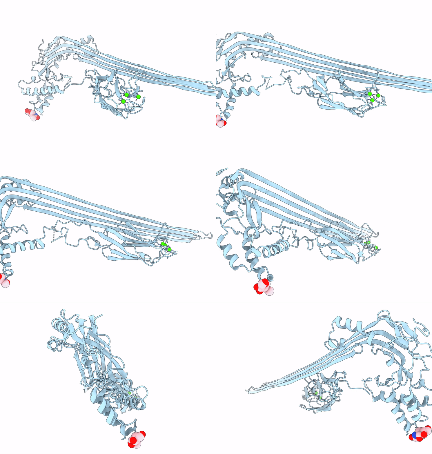
7PAG image
Deposition Date 2021-07-29
Release Date 2022-02-16
Last Version Date 2024-11-13
Entry Detail
PDB ID:
7PAG
Keywords:
Title:
The pore conformation of lymphocyte perforin
Biological Source:
Source Organism:
Mus musculus (Taxon ID: 10090)
Host Organism:
Method Details:
Experimental Method:
Resolution:
4.00 Å
Aggregation State:
PARTICLE
Reconstruction Method:
SINGLE PARTICLE
Macromolecular Entities
Structures with similar UniProt ID
Protein Blast
Polymer Type:polypeptide(L)
Molecule:Perforin-1
Gene (Uniprot):Prf1
Chain IDs:A
Chain Length:540
Number of Molecules:1
Biological Source:Mus musculus
Primary Citation
The pore conformation of lymphocyte perforin.
Sci Adv 8 eabk3147 eabk3147 (2022)
PMID: 35148176 DOI: 10.1126/sciadv.abk3147

Abstact

Perforin is a pore-forming protein that facilitates rapid killing of pathogen-infected or cancerous cells by the immune system. Perforin is released from cytotoxic lymphocytes, together with proapoptotic granzymes, to bind to a target cell membrane where it oligomerizes and forms pores. The pores allow granzyme entry, which rapidly triggers the apoptotic death of the target cell. Here, we present a 4-Å resolution cryo-electron microscopy structure of the perforin pore, revealing previously unidentified inter- and intramolecular interactions stabilizing the assembly. During pore formation, the helix-turn-helix motif moves away from the bend in the central β sheet to form an intermolecular contact. Cryo-electron tomography shows that prepores form on the membrane surface with minimal conformational changes. Our findings suggest the sequence of conformational changes underlying oligomerization and membrane insertion, and explain how several pathogenic mutations affect function.

Legend

Protein

Chemical

Disease

Primary Citation of related structures