7P8V image
Deposition Date 2021-07-23
Release Date 2022-06-29
Last Version Date 2024-07-17
Entry Detail
PDB ID:
7P8V
Title:
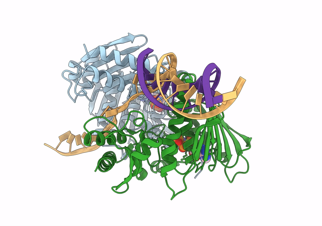
The structure of E. coli MutL bound to a 3' resected DNA end
Biological Source:
Source Organism:
Host Organism:
Method Details:
Experimental Method:
Resolution:
3.60 Å
Aggregation State:
PARTICLE
Reconstruction Method:
SINGLE PARTICLE
Macromolecular Entities
Polymer Type:polypeptide(L)
Molecule:DNA mismatch repair protein MutL
Gene (Uniprot):mutL
Chain IDs:A, B
Chain Length:615
Number of Molecules:2
Biological Source:Escherichia coli (strain K12)
Polymer Type:polydeoxyribonucleotide
Molecule:Template strand
Chain IDs:C
Chain Length:22
Number of Molecules:1
Biological Source:DNA molecule
Polymer Type:polydeoxyribonucleotide
Molecule:Primer strand
Chain IDs:D
Chain Length:13
Number of Molecules:1
Biological Source:DNA molecule
Primary Citation
MutL binds to 3' resected DNA ends and blocks DNA polymerase access.
Nucleic Acids Res. 50 6224 6234 (2022)
PMID: 35670670 DOI: 10.1093/nar/gkac432

Abstact

DNA mismatch repair removes mis-incorporated bases after DNA replication and reduces the error rate a 100-1000-fold. After recognition of a mismatch, a large section of up to a thousand nucleotides is removed from the daughter strand followed by re-synthesis. How these opposite activities are coordinated is poorly understood. Here we show that the Escherichia coli MutL protein binds to the 3' end of the resected strand and blocks access of Pol I and Pol III. The cryo-EM structure of an 85-kDa MutL-DNA complex, determined to 3.7 Å resolution, reveals a unique DNA binding mode that positions MutL at the 3' end of a primer-template, but not at a 5' resected DNA end or a blunt DNA end. Hence, our work reveals a novel role for MutL in the final stages of mismatch repair by preventing premature DNA synthesis during removal of the mismatched strand.

Legend

Protein

Chemical

Disease

Primary Citation of related structures