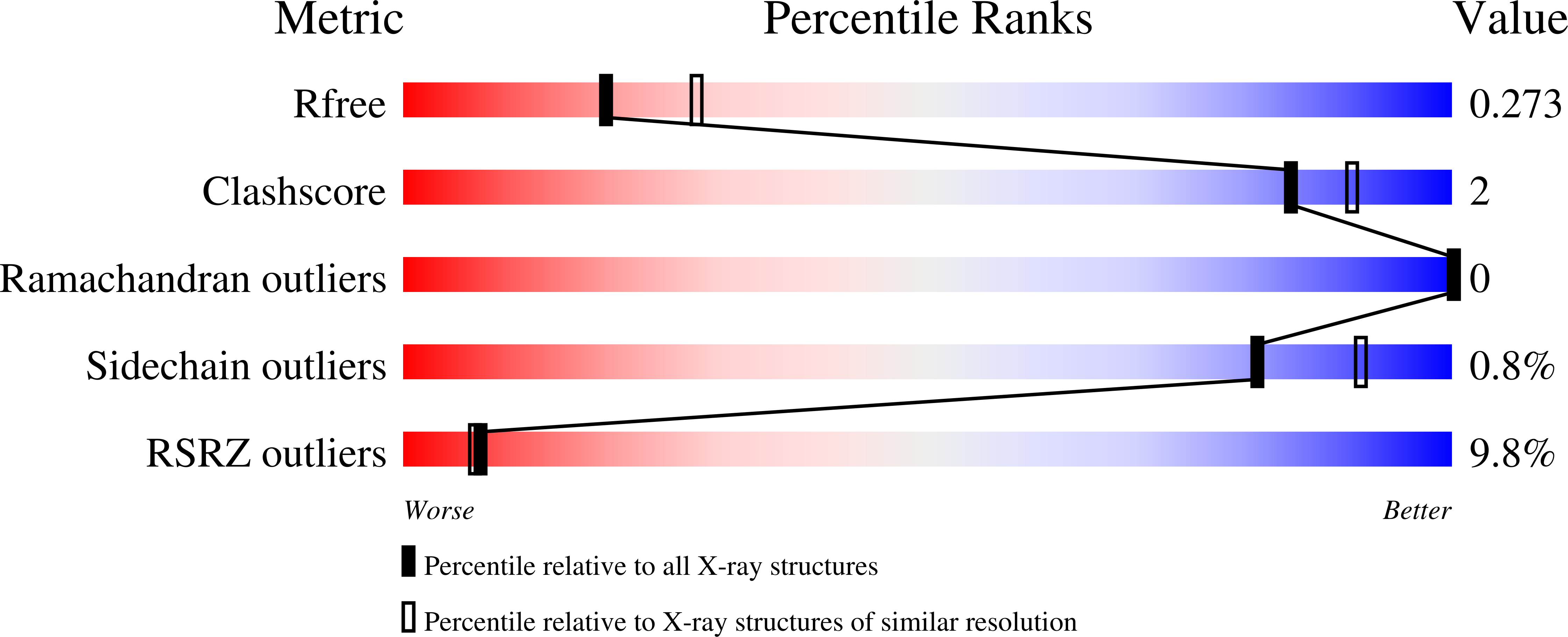
7P7H image
Entry Detail
PDB ID:
7P7H
Keywords:
Title:
Crystal structure of Casein Kinase I delta (CK1d) with alphaG-in conformation
Biological Source:
Source Organism:
PDB Version:
Deposition Date:
2021-07-19
Release Date:
2022-04-13
Method Details:
Experimental Method:
Resolution:
2.40 Å
R-Value Free:
0.26
R-Value Work:
0.22
R-Value Observed:
0.22
Space Group:
P 1 21 1
Macromolecular Entities
Polymer Type:polypeptide(L)
Description:Casein kinase I isoform delta
Chain IDs:A, B
Chain Length:296
Number of Molecules:2
Biological Source:Homo sapiens
Ligand Molecules
Primary Citation
Kinase domain autophosphorylation rewires the activity and substrate specificity of CK1 enzymes.
Mol.Cell 82 2006 ? (2022)
PMID: 35353987 DOI: 10.1016/j.molcel.2022.03.005

Abstact

CK1s are acidophilic serine/threonine kinases with multiple critical cellular functions; their misregulation contributes to cancer, neurodegenerative diseases, and sleep phase disorders. Here, we describe an evolutionarily conserved mechanism of CK1 activity: autophosphorylation of a threonine (T220 in human CK1δ) located at the N terminus of helix αG, proximal to the substrate binding cleft. Crystal structures and molecular dynamics simulations uncovered inherent plasticity in αG that increased upon T220 autophosphorylation. The phosphorylation-induced structural changes significantly altered the conformation of the substrate binding cleft, affecting substrate specificity. In T220 phosphorylated yeast and human CK1s, activity toward many substrates was decreased, but we also identified a high-affinity substrate that was phosphorylated more rapidly, and quantitative phosphoproteomics revealed that disrupting T220 autophosphorylation rewired CK1 signaling in Schizosaccharomyces pombe. T220 is present exclusively in the CK1 family, thus its autophosphorylation may have evolved as a unique regulatory mechanism for this important family.

Legend

Protein

Chemical

Disease

Primary Citation of related structures