7P6F image
Entry Detail
PDB ID:
7P6F
Title:
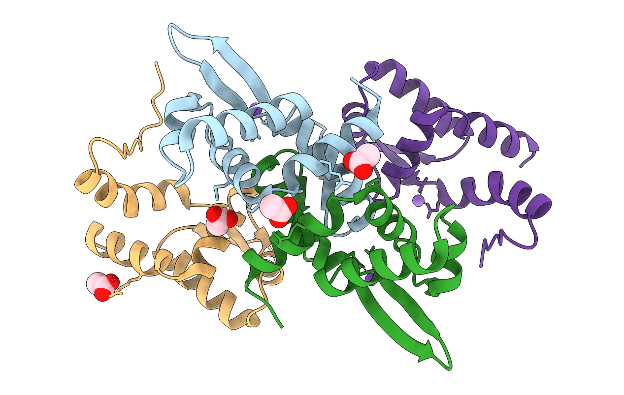
1.93 A resolution X-ray crystal structure of the transcriptional regulator SrnR from Streptomyces griseus
Biological Source:
Source Organism:
Host Organism:
PDB Version:
Deposition Date:
2021-07-16
Release Date:
2022-05-25
Method Details:
Experimental Method:
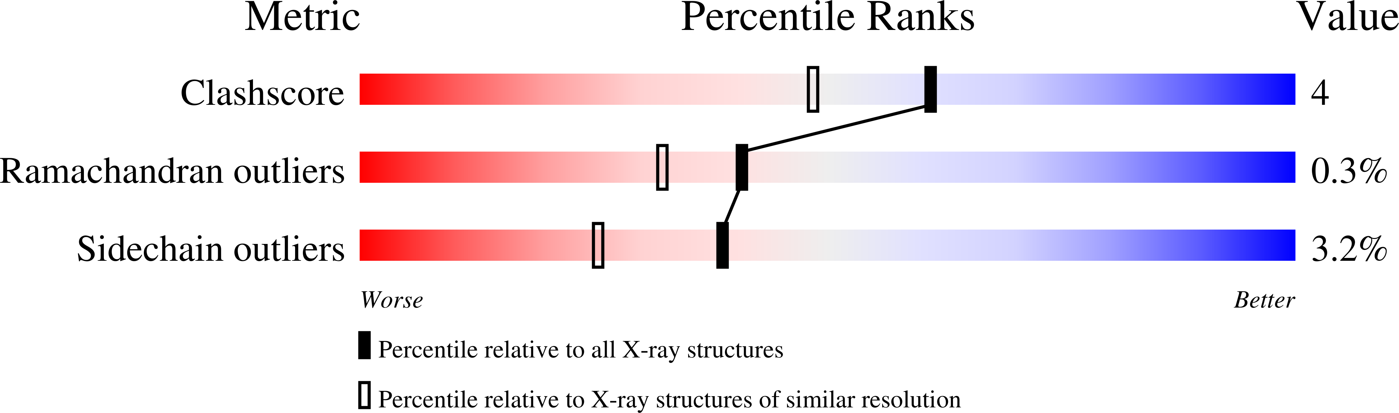
Resolution:
1.93 Å
R-Value Free:
0.21
R-Value Work:
0.17
Space Group:
P 62 2 2
Macromolecular Entities
Polymer Type:polypeptide(L)
Description:Transcriptional regulator SrnR
Chain IDs:A (auth: AAA), B (auth: BBB), C (auth: CCC), D (auth: DDD)
Chain Length:117
Number of Molecules:4
Biological Source:Streptomyces griseus
Primary Citation
Structure, dynamics, and function of SrnR, a transcription factor for nickel-dependent gene expression.
Metallomics 13 ? ? (2021)
PMID: 34850061 DOI: 10.1093/mtomcs/mfab069

Abstact

Streptomyces griseus, a bacterium producing antibacterial drugs and featuring possible application in phytoremediation, expresses two metal-dependent superoxide dismutase (SOD) enzymes, containing either Fe(II) or Ni(II) in their active site. In particular, the alternative expression of the two proteins occurs in a metal-dependent mode, with the Fe(II)-enzyme gene (sodF) repressed at high intracellular Ni(II) concentrations by a two-component system (TCS). This complex involves two proteins, namely SgSrnR and SgSrnQ, which represent the transcriptional regulator and the Ni(II) sensor of the system, respectively. SgSrnR belongs to the ArsR/SmtB family of metal-dependent transcription factors; in the apo-form and in the absence of SgSrnQ, it can bind the DNA operator of sodF, upregulating gene transcription. According to a recently proposed hypothesis, Ni(II) binding to SgSrnQ would promote its interaction with SgSrnR, causing the release of the complex from DNA and the consequent downregulation of the sodF expression. SgSrnQ is predicted to be highly disordered, thus the understanding, at the molecular level, of how the SgSrnR/SgSrnQ TCS specifically responds to Ni(II) requires the knowledge of the structural, dynamic, and functional features of SgSrnR. These were investigated synergistically in this work using X-ray crystallography, nuclear magnetic resonance (NMR) spectroscopy, atomistic molecular dynamics calculations, isothermal titration calorimetry, and in silico molecular docking. The results reveal that the homodimeric apo-SgSrnR binds to its operator in a two-step process that involves the more rigid globular portion of the protein and leaves its largely disordered regions available to possibly interact with the disordered SgSrnQ in a Ni-dependent process.

Legend

Protein

Chemical

Disease

Primary Citation of related structures