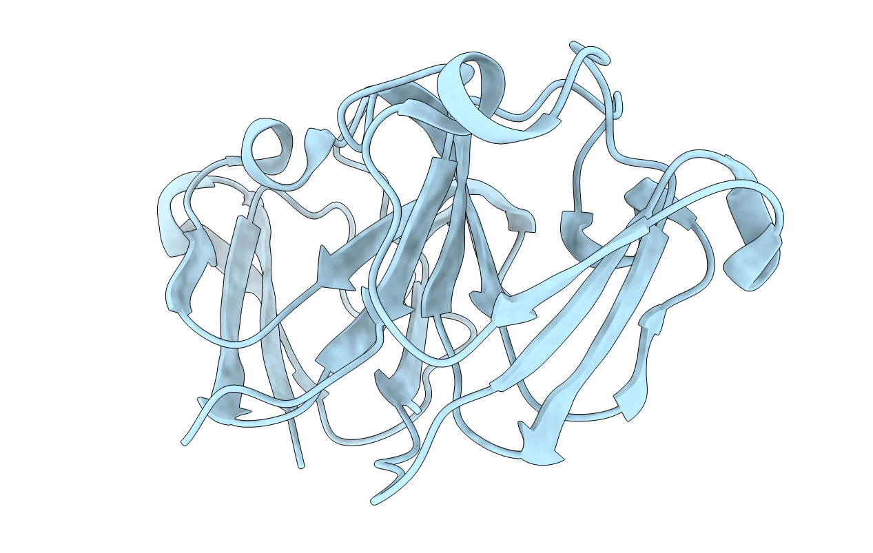
7P53 image
Entry Detail
PDB ID:
7P53
Title:
Crystal Structure of Human gamma-D-crystallin mutant C110M at 1.57 Angstroms resolution
Biological Source:
Source Organism:
PDB Version:
Deposition Date:
2021-07-14
Release Date:
2021-10-13
Method Details:
Experimental Method:
Resolution:
1.57 Å
R-Value Free:
0.27
R-Value Work:
0.21
R-Value Observed:
0.21
Space Group:
P 21 21 21
Macromolecular Entities
Polymer Type:polypeptide(L)
Description:Gamma-crystallin D
Mutations:C110M
Chain IDs:A (auth: X)
Chain Length:173
Number of Molecules:1
Biological Source:Homo sapiens
Primary Citation
Surface Exposed Free Cysteine Suppresses Crystallization of Human gamma D-Crystallin.
J.Mol.Biol. 433 167252 167252 (2021)
PMID: 34537240 DOI: 10.1016/j.jmb.2021.167252

Abstact

Human γD-crystallin (HGD) has remarkable stability against condensation in the human lens, sometimes over a whole lifetime. The native protein has a surface exposed free cysteine that forms dimers (Benedek, 1997; Ramkumar et al., 1864)1,2 without specific biological function and leads to further protein association and/or aggregation, which creates a paradox for understanding its stability. Previous work has demonstrated that chemical modification of the protein at the free cysteine (C110), increases the temperature at which liquid-liquid phase separation occurs (LLPS), lowers protein solubility and suggests an important role for this amino acid in maintaining its long-term resistance to condensation. Here we demonstrate that mutation of the cysteine does not alter the structure or solubility (liquidus) line for the protein, but dramatically increases the protein crystal nucleation rate following LLPS, suggesting that the free cysteine has a vital role in suppressing crystallization in the human lens.

Legend

Protein

Chemical

Disease

Primary Citation of related structures