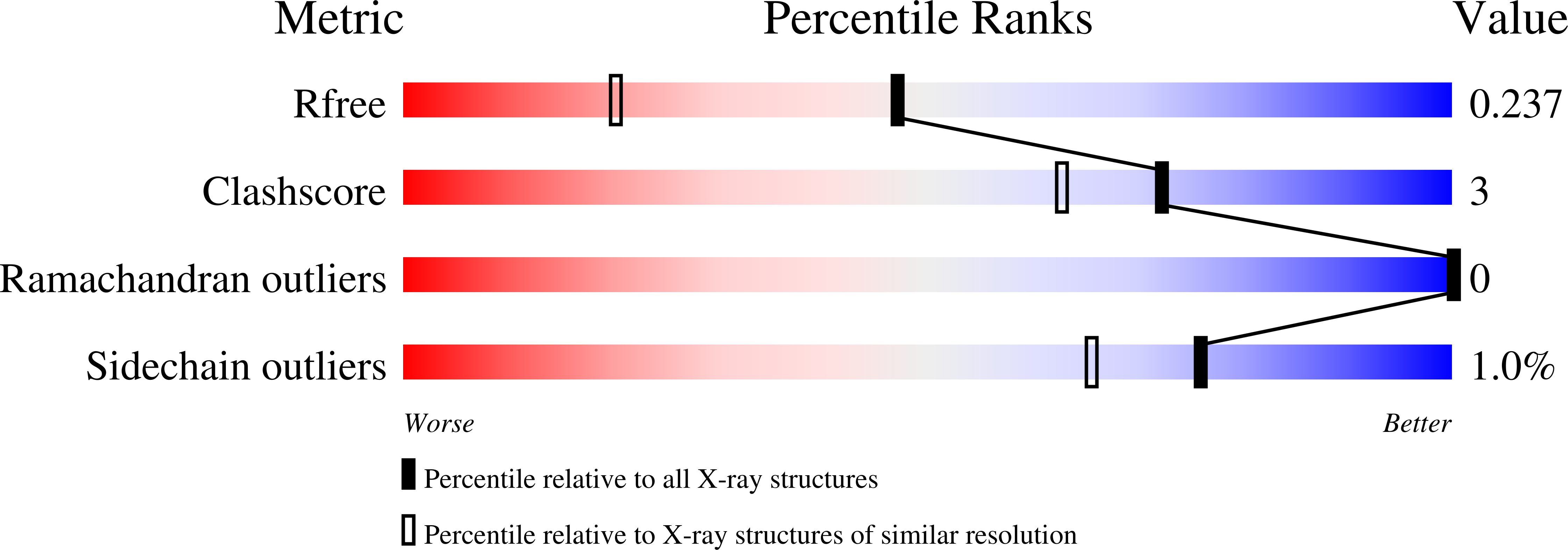
7P4B image
Deposition Date 2021-07-10
Release Date 2022-07-27
Last Version Date 2024-10-09
Entry Detail
PDB ID:
7P4B
Keywords:
Title:
HLA-E*01:03 in complex with IL9
Biological Source:
Source Organism:
Host Organism:
Method Details:
Experimental Method:
Resolution:
1.72 Å
R-Value Free:
0.23
R-Value Work:
0.20
R-Value Observed:
0.20
Space Group:
C 1 2 1
Macromolecular Entities
Polymer Type:polypeptide(L)
Molecule:HLA class I histocompatibility antigen, alpha chain E
Gene (Uniprot):HLA-E
Chain IDs:A, C, E, G
Chain Length:277
Number of Molecules:4
Biological Source:Homo sapiens
Polymer Type:polypeptide(L)
Molecule:Beta-2-microglobulin
Gene (Uniprot):B2M
Chain IDs:B, D, F, H
Chain Length:100
Number of Molecules:4
Biological Source:Homo sapiens
Polymer Type:polypeptide(L)
Molecule:ESAT-6-like protein EsxH
Gene (Uniprot):esxH
Chain IDs:I (auth: P), J (auth: Q), K (auth: R), L (auth: Z)
Chain Length:9
Number of Molecules:4
Biological Source:Mycobacterium tuberculosis H37Rv
Primary Citation
Primary and secondary functions of HLA-E are determined by stability and conformation of the peptide-bound complexes.
Cell Rep 39 110959 110959 (2022)
PMID: 35705051 DOI: 10.1016/j.celrep.2022.110959

Abstact

MHC-E regulates NK cells by displaying MHC class Ia signal peptides (VL9) to NKG2A:CD94 receptors. MHC-E can also present sequence-diverse, lower-affinity, pathogen-derived peptides to T cell receptors (TCRs) on CD8+ T cells. To understand these affinity differences, human MHC-E (HLA-E)-VL9 versus pathogen-derived peptide structures are compared. Small-angle X-ray scatter (SAXS) measures biophysical parameters in solution, allowing comparison with crystal structures. For HLA-E-VL9, there is concordance between SAXS and crystal parameters. In contrast, HLA-E-bound pathogen-derived peptides produce larger SAXS dimensions that reduce to their crystallographic dimensions only when excess peptide is supplied. Further crystallographic analysis demonstrates three amino acids, exclusive to MHC-E, that not only position VL9 close to the α2 helix, but also allow non-VL9 peptide binding with re-configuration of a key TCR-interacting α2 region. Thus, non-VL9-bound peptides introduce an alternative peptide-binding motif and surface recognition landscape, providing a likely basis for VL9- and non-VL9-HLA-E immune discrimination.

Legend

Protein

Chemical

Disease

Primary Citation of related structures