Deposition Date
2021-07-09
Release Date
2021-10-06
Last Version Date
2024-01-31
Entry Detail
PDB ID:
7P46
Keywords:
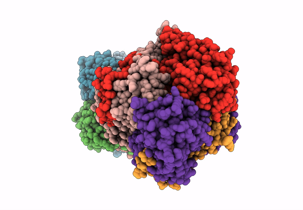
Title:
Crystal Structure of Xanthomonas campestris Tryptophan 2,3-dioxygenase (TDO)
Biological Source:
Source Organism(s):
Expression System(s):
Method Details:
Experimental Method:
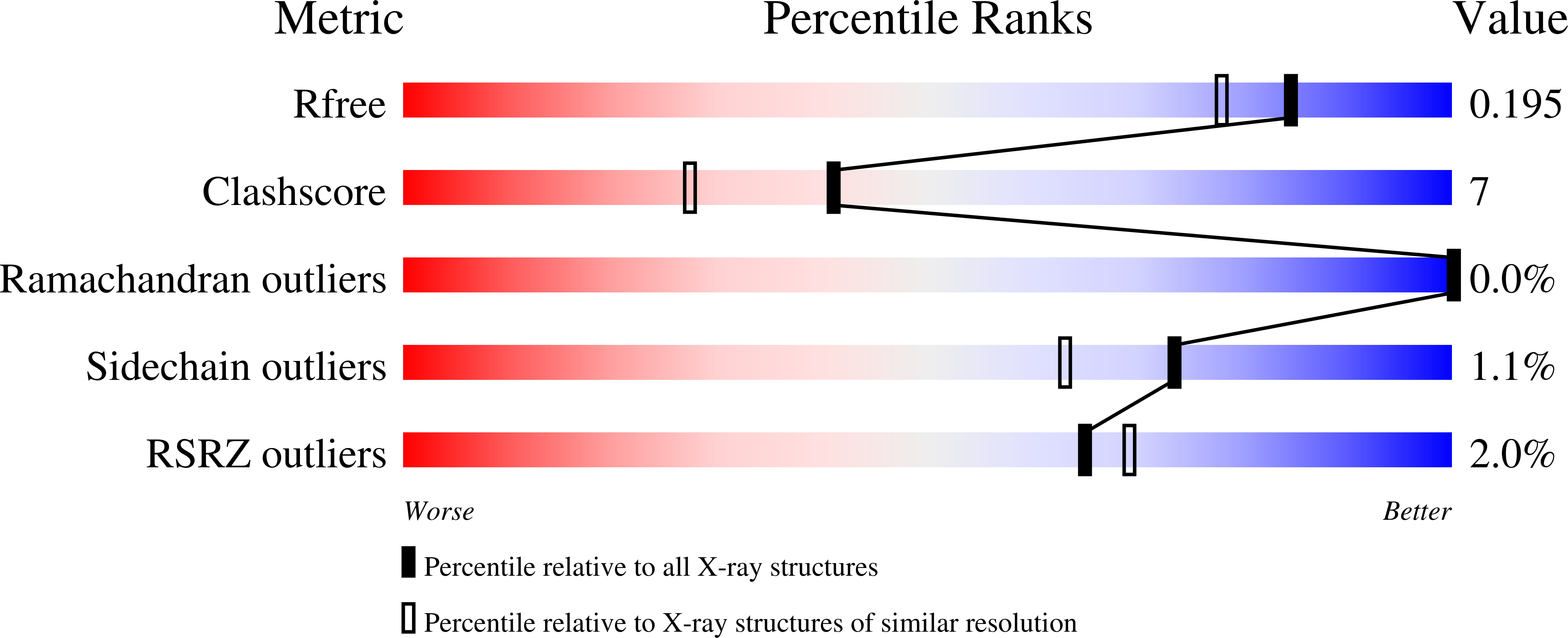
Resolution:
1.70 Å
R-Value Free:
0.18
R-Value Work:
0.14
R-Value Observed:
0.15
Space Group:
P 1 21 1