Deposition Date
2021-07-08
Release Date
2022-02-23
Last Version Date
2024-07-17
Entry Detail
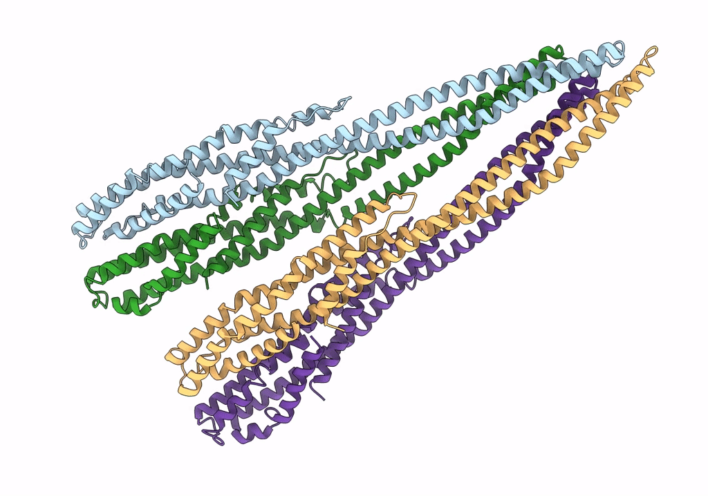
Biological Source:
Source Organism(s):
Vibrio cholerae O1 biovar El Tor str. N16961 (Taxon ID: 243277)
Expression System(s):
Method Details:
Experimental Method:
Resolution:
3.65 Å
Aggregation State:
HELICAL ARRAY
Reconstruction Method:
HELICAL