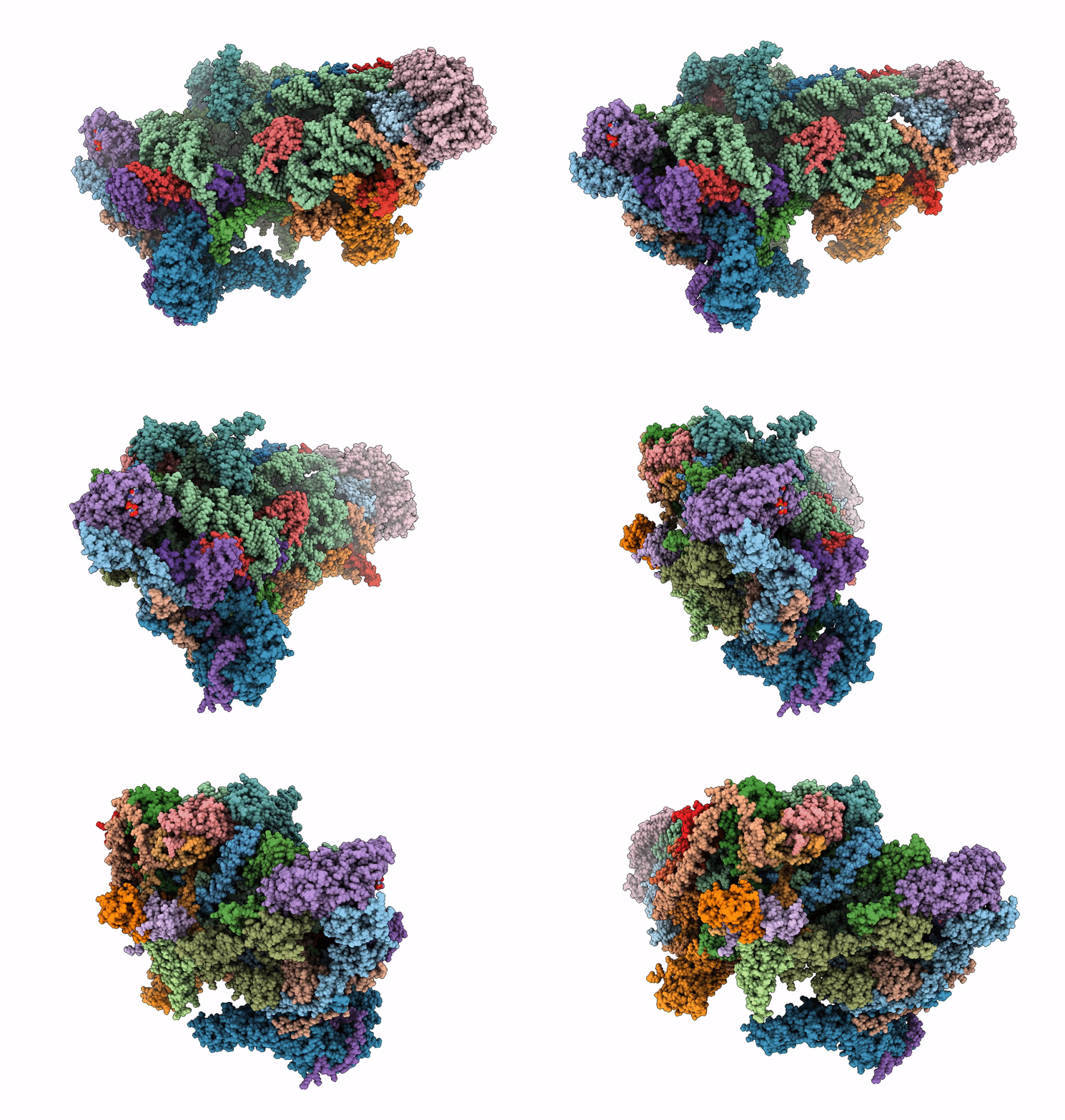
7P2E image
Entry Detail
PDB ID:
7P2E
EMDB ID:
Keywords:
Title:
Human mitochondrial ribosome small subunit in complex with IF3, GMPPMP and streptomycin
Biological Source:
Source Organism:
Host Organism:
PDB Version:
Deposition Date:
2021-07-05
Release Date:
2022-07-27
Method Details:
Experimental Method:
Resolution:
2.40 Å
Aggregation State:
PARTICLE
Reconstruction Method:
SINGLE PARTICLE
Macromolecular Entities
Polymer Type:polypeptide(L)
Description:28S ribosomal protein S34, mitochondrial
Chain IDs:AA (auth: 0)
Chain Length:218
Number of Molecules:1
Biological Source:Homo sapiens
Polymer Type:polypeptide(L)
Description:28S ribosomal protein S35, mitochondrial
Chain IDs:BA (auth: 1)
Chain Length:323
Number of Molecules:1
Biological Source:Homo sapiens
Polymer Type:polypeptide(L)
Description:Coiled-coil-helix-coiled-coil-helix domain-containing protein 1
Chain IDs:CA (auth: 2)
Chain Length:117
Number of Molecules:1
Biological Source:Homo sapiens
Polymer Type:polypeptide(L)
Description:Aurora kinase A-interacting protein
Chain IDs:DA (auth: 3)
Chain Length:199
Number of Molecules:1
Biological Source:Homo sapiens
Polymer Type:polypeptide(L)
Description:Pentatricopeptide repeat domain-containing protein 3, mitochondrial
Chain IDs:EA (auth: 4)
Chain Length:689
Number of Molecules:1
Biological Source:Homo sapiens
Polymer Type:polypeptide(L)
Description:Translation initiation factor IF-3, mitochondrial
Chain IDs:FA (auth: 8)
Chain Length:285
Number of Molecules:1
Biological Source:Homo sapiens
Polymer Type:polyribonucleotide
Description:12S mitochondrial rRNA
Chain IDs:A
Chain Length:955
Number of Molecules:1
Biological Source:Homo sapiens
Polymer Type:polypeptide(L)
Description:28S ribosomal protein S2, mitochondrial
Chain IDs:B
Chain Length:296
Number of Molecules:1
Biological Source:Homo sapiens
Polymer Type:polypeptide(L)
Description:28S ribosomal protein S24, mitochondrial
Chain IDs:C
Chain Length:167
Number of Molecules:1
Biological Source:Homo sapiens
Polymer Type:polypeptide(L)
Description:28S ribosomal protein S5, mitochondrial
Chain IDs:D
Chain Length:430
Number of Molecules:1
Biological Source:Homo sapiens
Polymer Type:polypeptide(L)
Description:28S ribosomal protein S6, mitochondrial
Chain IDs:E
Chain Length:125
Number of Molecules:1
Biological Source:Homo sapiens
Polymer Type:polypeptide(L)
Description:28S ribosomal protein S7, mitochondrial
Chain IDs:F
Chain Length:242
Number of Molecules:1
Biological Source:Homo sapiens
Polymer Type:polypeptide(L)
Description:28S ribosomal protein S9, mitochondrial
Chain IDs:G
Chain Length:396
Number of Molecules:1
Biological Source:Homo sapiens
Polymer Type:polypeptide(L)
Description:28S ribosomal protein S10, mitochondrial
Chain IDs:H
Chain Length:201
Number of Molecules:1
Biological Source:Homo sapiens
Polymer Type:polypeptide(L)
Description:28S ribosomal protein S11, mitochondrial
Chain IDs:I
Chain Length:194
Number of Molecules:1
Biological Source:Homo sapiens
Polymer Type:polypeptide(L)
Description:28S ribosomal protein S12, mitochondrial
Chain IDs:J
Chain Length:138
Number of Molecules:1
Biological Source:Homo sapiens
Polymer Type:polypeptide(L)
Description:28S ribosomal protein S14, mitochondrial
Chain IDs:K
Chain Length:128
Number of Molecules:1
Biological Source:Homo sapiens
Polymer Type:polypeptide(L)
Description:28S ribosomal protein S15, mitochondrial
Chain IDs:L
Chain Length:257
Number of Molecules:1
Biological Source:Homo sapiens
Polymer Type:polypeptide(L)
Description:28S ribosomal protein S16, mitochondrial
Chain IDs:M
Chain Length:137
Number of Molecules:1
Biological Source:Homo sapiens
Polymer Type:polypeptide(L)
Description:28S ribosomal protein S17, mitochondrial
Chain IDs:N
Chain Length:130
Number of Molecules:1
Biological Source:Homo sapiens
Polymer Type:polypeptide(L)
Description:28S ribosomal protein S18b, mitochondrial
Chain IDs:O
Chain Length:258
Number of Molecules:1
Biological Source:Homo sapiens
Polymer Type:polypeptide(L)
Description:28S ribosomal protein S18c, mitochondrial
Chain IDs:P
Chain Length:142
Number of Molecules:1
Biological Source:Homo sapiens
Polymer Type:polypeptide(L)
Description:28S ribosomal protein S21, mitochondrial
Chain IDs:Q
Chain Length:86
Number of Molecules:1
Biological Source:Homo sapiens
Polymer Type:polypeptide(L)
Description:28S ribosomal protein S22, mitochondrial
Chain IDs:R
Chain Length:360
Number of Molecules:1
Biological Source:Homo sapiens
Polymer Type:polypeptide(L)
Description:28S ribosomal protein S23, mitochondrial
Chain IDs:S
Chain Length:190
Number of Molecules:1
Biological Source:Homo sapiens
Polymer Type:polypeptide(L)
Description:28S ribosomal protein S25, mitochondrial
Chain IDs:T
Chain Length:173
Number of Molecules:1
Biological Source:Homo sapiens
Polymer Type:polypeptide(L)
Description:28S ribosomal protein S26, mitochondrial
Chain IDs:U
Chain Length:205
Number of Molecules:1
Biological Source:Homo sapiens
Polymer Type:polypeptide(L)
Description:28S ribosomal protein S27, mitochondrial
Chain IDs:V
Chain Length:414
Number of Molecules:1
Biological Source:Homo sapiens
Polymer Type:polypeptide(L)
Description:28S ribosomal protein S28, mitochondrial
Chain IDs:W
Chain Length:187
Number of Molecules:1
Biological Source:Homo sapiens
Polymer Type:polypeptide(L)
Description:28S ribosomal protein S29, mitochondrial
Chain IDs:X
Chain Length:398
Number of Molecules:1
Biological Source:Homo sapiens
Polymer Type:polypeptide(L)
Description:28S ribosomal protein S31, mitochondrial
Chain IDs:Y
Chain Length:395
Number of Molecules:1
Biological Source:Homo sapiens
Polymer Type:polypeptide(L)
Description:28S ribosomal protein S33, mitochondrial
Chain IDs:Z
Chain Length:106
Number of Molecules:1
Biological Source:Homo sapiens
Modified Residue
Compound ID Chain ID Parent Comp ID Details 2D Image
AYA Q ALA modified residue
Primary Citation
Structure of the mitoribosomal small subunit with streptomycin reveals Fe-S clusters and physiological molecules.
Elife 11 ? ? (2022)
PMID: 36480258 DOI: 10.7554/eLife.77460

Abstact

The mitoribosome regulates cellular energy production, and its dysfunction is associated with aging. Inhibition of the mitoribosome can be caused by off-target binding of antimicrobial drugs and was shown to be coupled with a bilateral decreased visual acuity. Previously, we reported mitochondria-specific protein aspects of the mitoribosome, and in this article we present a 2.4-Å resolution structure of the small subunit in a complex with the anti-tuberculosis drug streptomycin that reveals roles of non-protein components. We found iron-sulfur clusters that are coordinated by different mitoribosomal proteins, nicotinamide adenine dinucleotide (NAD) associated with rRNA insertion, and posttranslational modifications. This is the first evidence of inter-protein coordination of iron-sulfur, and the finding of iron-sulfur clusters and NAD as fundamental building blocks of the mitoribosome directly links to mitochondrial disease and aging. We also report details of streptomycin interactions, suggesting that the mitoribosome-bound streptomycin is likely to be in hydrated gem-diol form and can be subjected to other modifications by the cellular milieu. The presented approach of adding antibiotics to cultured cells can be used to define their native structures in a bound form under more physiological conditions, and since streptomycin is a widely used drug for treatment, the newly resolved features can serve as determinants for targeting.

Legend

Protein

Chemical

Disease

Primary Citation of related structures