7P2B image
Deposition Date 2021-07-05
Release Date 2022-05-18
Last Version Date 2024-10-16
Entry Detail
PDB ID:
7P2B
Title:
Crystal structure of human gelsolin amyloid mutant A551P
Biological Source:
Source Organism:
Homo sapiens (Taxon ID: 9606)
Host Organism:
Method Details:
Experimental Method:
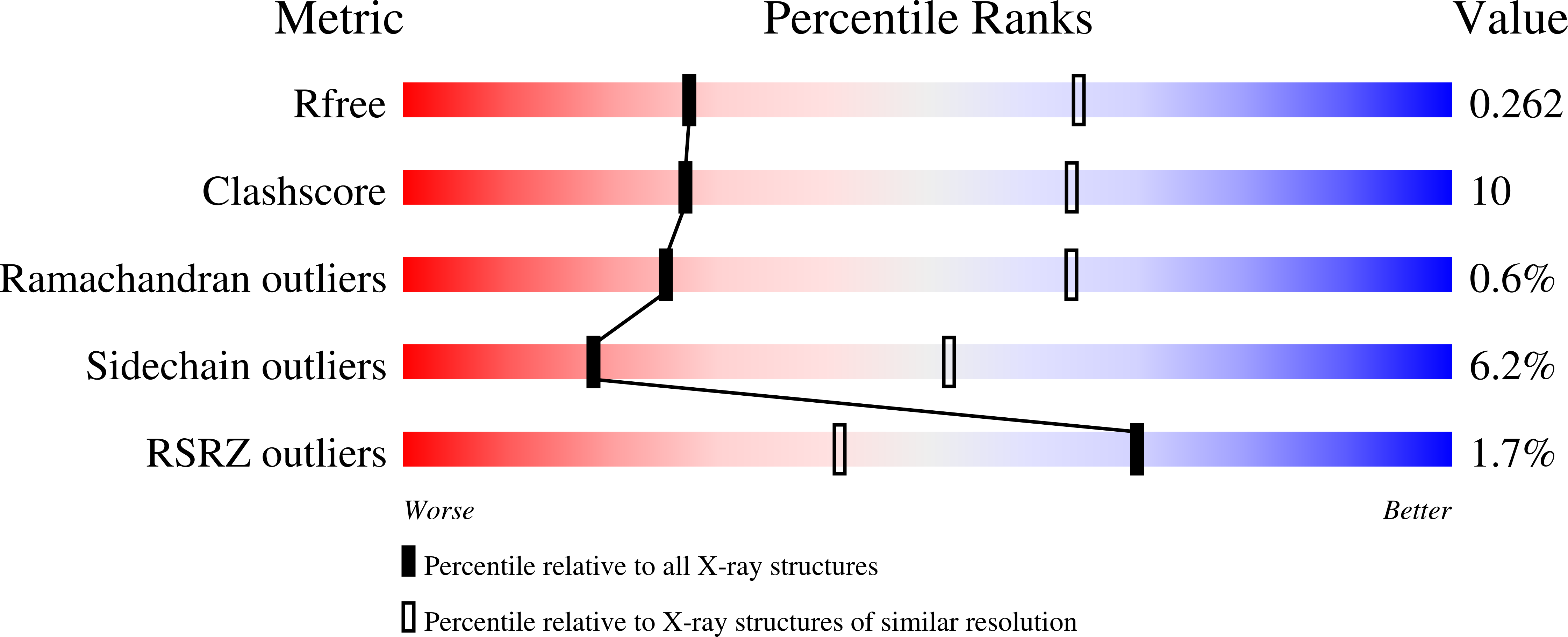
Resolution:
3.00 Å
R-Value Free:
0.26
R-Value Work:
0.21
R-Value Observed:
0.21
Space Group:
P 4 21 2
Macromolecular Entities
Structures with similar UniProt ID
Protein Blast
Polymer Type:polypeptide(L)
Molecule:Gelsolin
Gene (Uniprot):GSN
Mutagens:A551P
Chain IDs:A, B
Chain Length:778
Number of Molecules:2
Biological Source:Homo sapiens
Primary Citation
A novel hotspot of gelsolin instability triggers an alternative mechanism of amyloid aggregation.
Comput Struct Biotechnol J 19 6355 6365 (2021)
PMID: 34938411 DOI: 10.1016/j.csbj.2021.11.025

Abstact

Gelsolin comprises six homologous domains, named G1 to G6. Single point substitutions in this protein are responsible for AGel amyloidosis, a hereditary disease causing progressive corneal lattice dystrophy, cutis laxa, and polyneuropathy. Although several different amyloidogenic variants of gelsolin have been identified, only the most common mutants present in the G2 domain have been thoroughly characterized, leading to clarification of the functional mechanism. The molecular events underlying the pathological aggregation of 3 recently identified mutations, namely A551P, E553K and M517R, all localized at the interface between G4 and G5, are here explored for the first time. Structural studies point to destabilization of the interface between G4 and G5 due to three structural determinants: β-strand breaking, steric hindrance and/or charge repulsion, all implying impairment of interdomain contacts. Such rearrangements decrease the temperature and pressure stability of gelsolin but do not alter its susceptibility to furin cleavage, the first event in the canonical aggregation pathway. These variants also have a greater tendency to aggregate in the unproteolysed forms and exhibit higher proteotoxicity in a C. elegans-based assay. Our data suggest that aggregation of G4G5 variants follows an alternative, likely proteolysis-independent, pathway.

Legend

Protein

Chemical

Disease

Primary Citation of related structures
Feedback Form
Name
Email
Institute
Feedback