7P1H image
Entry Detail
PDB ID:
7P1H
EMDB ID:
Keywords:
Title:
Structure of the V. vulnificus ExoY-G-actin-profilin complex
Biological Source:
PDB Version:
Deposition Date:
2021-07-01
Release Date:
2021-11-17
Method Details:
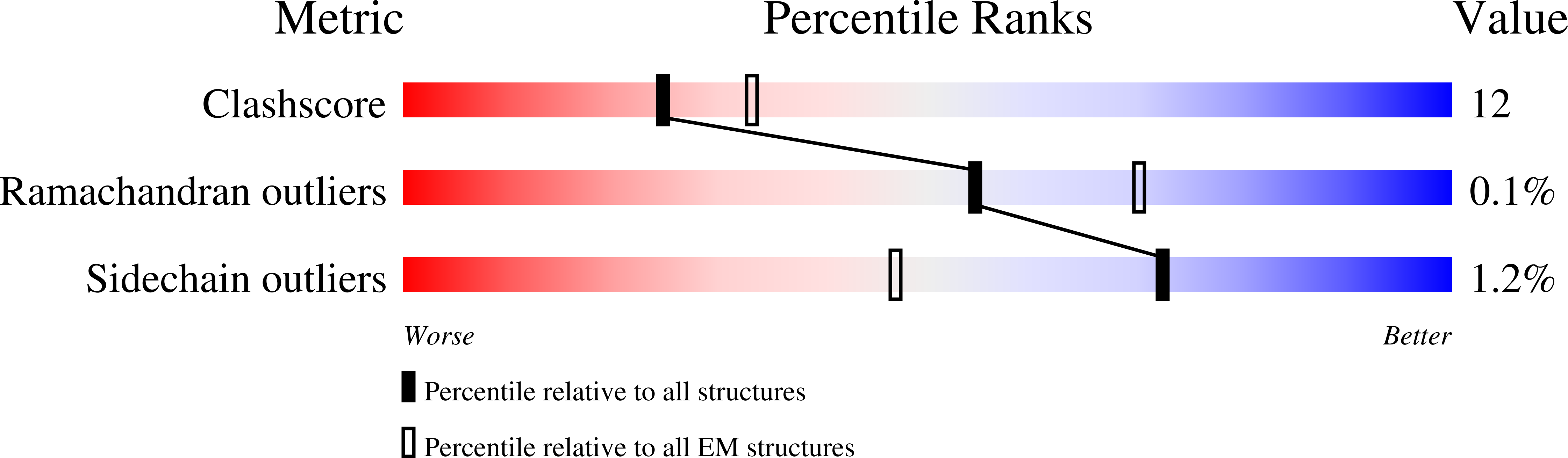
Experimental Method:
Resolution:
3.90 Å
Aggregation State:
PARTICLE
Reconstruction Method:
SINGLE PARTICLE
Macromolecular Entities
Polymer Type:polypeptide(L)
Description:Maltose/maltodextrin-binding periplasmic protein,RTX-toxin
Chain IDs:A
Chain Length:827
Number of Molecules:1
Biological Source:Escherichia coli (strain K12), Vibrio vulnificus
Polymer Type:polypeptide(L)
Description:Actin, cytoplasmic 1
Mutations:C272A
Chain IDs:B
Chain Length:372
Number of Molecules:1
Biological Source:Homo sapiens
Polymer Type:polypeptide(L)
Description:Profilin-1
Chain IDs:C (auth: P)
Chain Length:140
Number of Molecules:1
Biological Source:Homo sapiens
Modified Residue
Compound ID Chain ID Parent Comp ID Details 2D Image
HIC B HIS modified residue
Primary Citation
Mechanism of actin-dependent activation of nucleotidyl cyclase toxins from bacterial human pathogens.
Nat Commun 12 6628 6628 (2021)
PMID: 34785651 DOI: 10.1038/s41467-021-26889-2

Abstact

Bacterial human pathogens secrete initially inactive nucleotidyl cyclases that become potent enzymes by binding to actin inside eukaryotic host cells. The underlying molecular mechanism of this activation is, however, unclear. Here, we report structures of ExoY from Pseudomonas aeruginosa and Vibrio vulnificus bound to their corresponding activators F-actin and profilin-G-actin. The structures reveal that in contrast to the apo-state, two flexible regions become ordered and interact strongly with actin. The specific stabilization of these regions results in an allosteric stabilization of the nucleotide binding pocket and thereby to an activation of the enzyme. Differences in the sequence and conformation of the actin-binding regions are responsible for the selective binding to either F- or G-actin. Other nucleotidyl cyclase toxins that bind to calmodulin rather than actin undergo a similar disordered-to-ordered transition during activation, suggesting that the allosteric activation-by-stabilization mechanism of ExoY is conserved in these enzymes, albeit the different activator.

Legend

Protein

Chemical

Disease

Primary Citation of related structures