Deposition Date
2021-07-01
Release Date
2021-11-17
Last Version Date
2021-12-01
Entry Detail
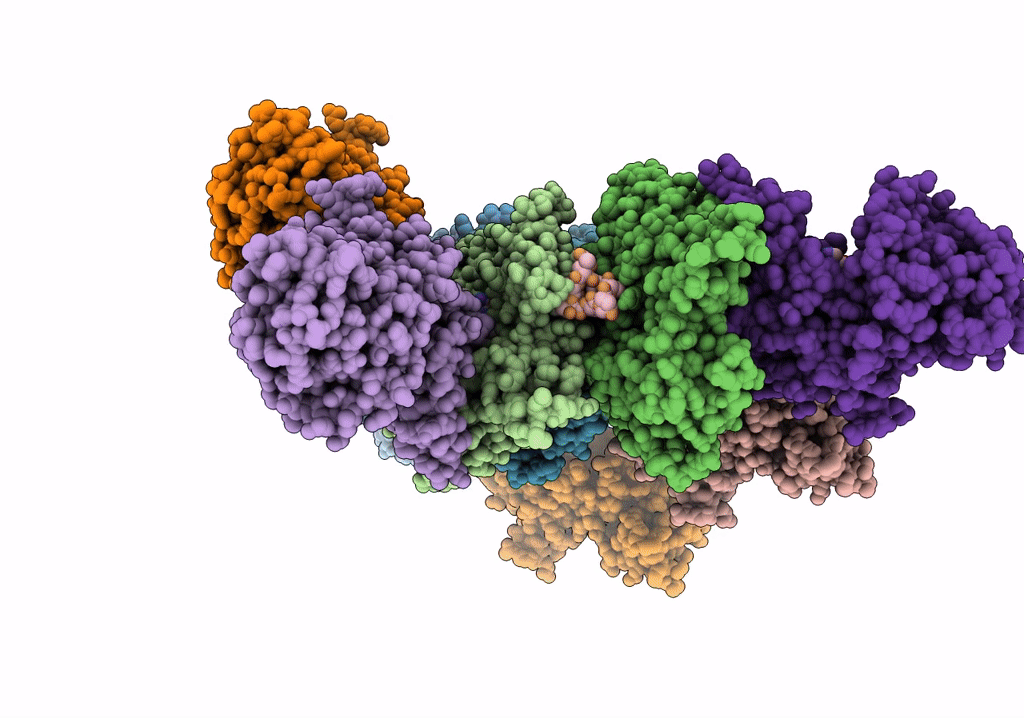
Biological Source:
Source Organism(s):
Escherichia coli K-12 (Taxon ID: 83333)
Pseudomonas aeruginosa PAO1 (Taxon ID: 208964)
Amanita phalloides (Taxon ID: 67723)
Oryctolagus cuniculus (Taxon ID: 9986)
Pseudomonas aeruginosa PAO1 (Taxon ID: 208964)
Amanita phalloides (Taxon ID: 67723)
Oryctolagus cuniculus (Taxon ID: 9986)
Expression System(s):
Method Details:
Experimental Method:
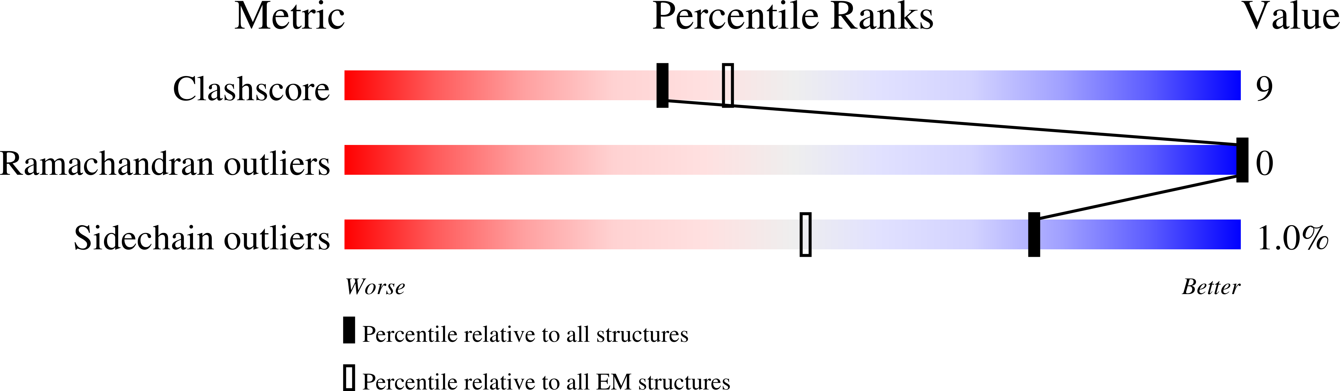
Resolution:
3.20 Å
Aggregation State:
FILAMENT
Reconstruction Method:
SINGLE PARTICLE