
Deposition Date
2021-06-30
Release Date
2022-03-23
Last Version Date
2024-11-20
Entry Detail
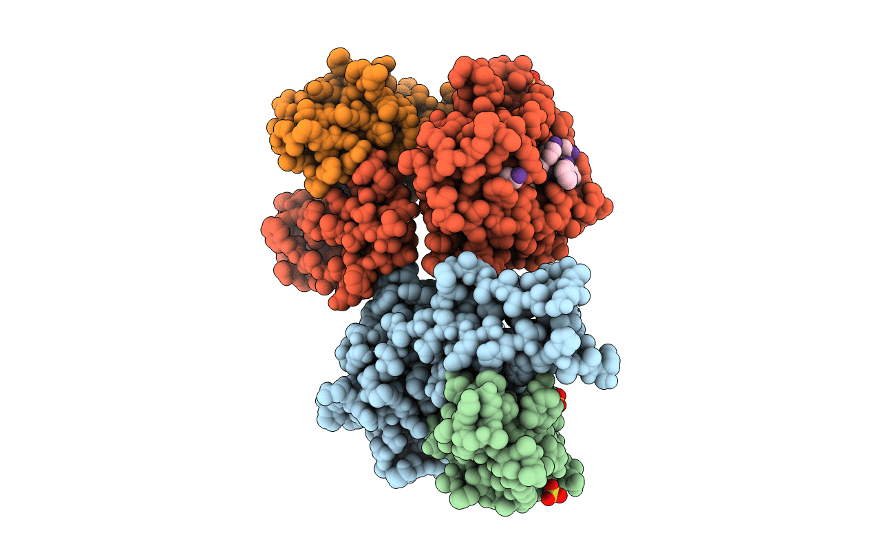
PDB ID:
7P0T
Keywords:
Title:
CRYSTAL STRUCTURE OF THE MURINE CLASS I MAJOR HISTOCOMPATIBILITY COMPLEX H-2DB IN COMPLEX WITH LCMV-DERIVED GP33 PEPTIDE with D-AMINOACID
Biological Source:
Source Organism(s):
Mus musculus (Taxon ID: 10090)
Lymphocytic choriomeningitis mammarenavirus (Taxon ID: 11623)
Lymphocytic choriomeningitis mammarenavirus (Taxon ID: 11623)
Expression System(s):
Method Details:
Experimental Method:
Resolution:
2.61 Å
R-Value Free:
0.24
R-Value Work:
0.19
R-Value Observed:
0.20
Space Group:
C 1 2 1
