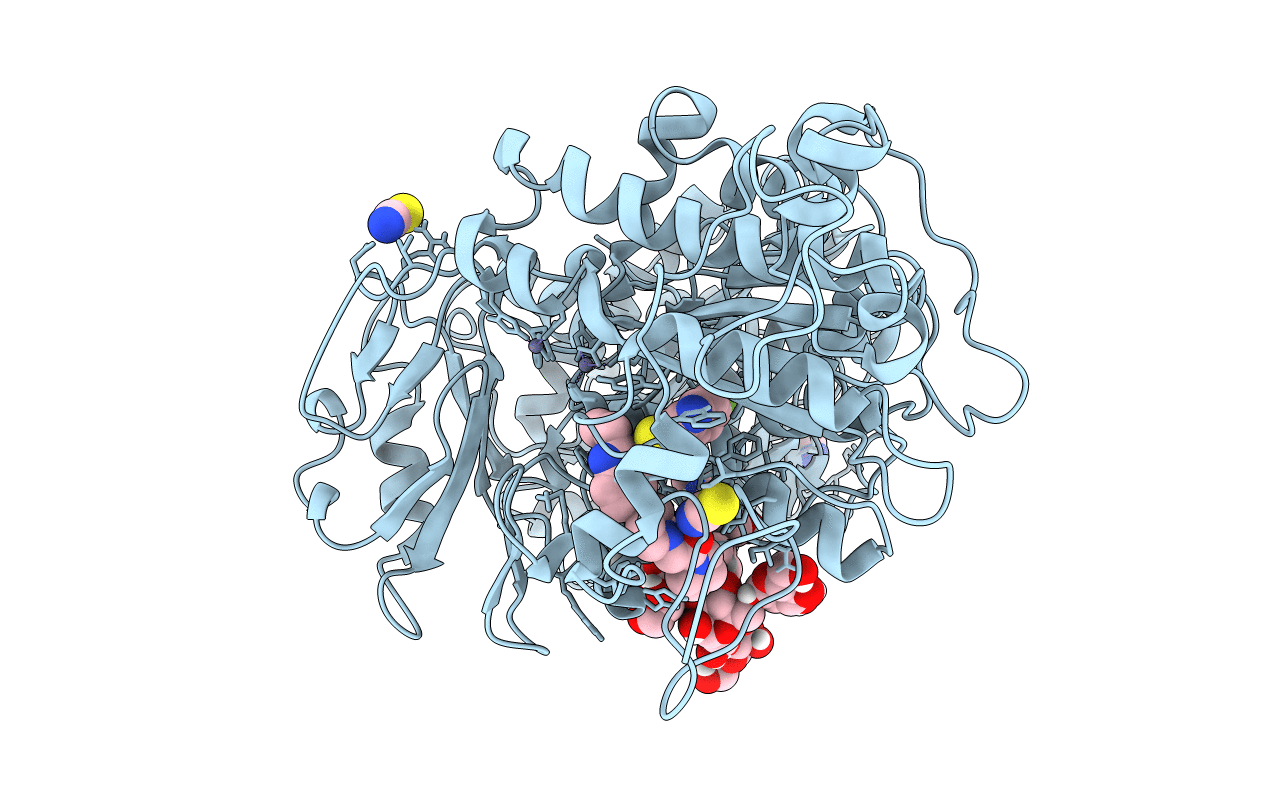
7P0K image
Deposition Date 2021-06-29
Release Date 2022-07-13
Last Version Date 2024-10-16
Entry Detail
PDB ID:
7P0K
Keywords:
Title:
Crystal structure of Autotaxin (ENPP2) with 18F-labeled positron emission tomography ligand
Biological Source:
Source Organism:
Host Organism:
Method Details:
Experimental Method:
Resolution:
2.20 Å
R-Value Free:
0.27
R-Value Work:
0.21
Space Group:
P 21 21 21
Macromolecular Entities
Polymer Type:polypeptide(L)
Molecule:Isoform 2 of Ectonucleotide pyrophosphatase/phosphodiesterase family member 2
Gene (Uniprot):Enpp2
Mutations:N53A,N410A,N806A
Chain IDs:A (auth: AAA)
Chain Length:770
Number of Molecules:1
Biological Source:Rattus norvegicus
Primary Citation
Imaging Autotaxin In Vivo with 18 F-Labeled Positron Emission Tomography Ligands
J Med Chem 64 15053 15068 (2021)
PMID: 34662125 DOI: 10.1021/acs.jmedchem.1c00913

Abstact

Autotaxin (ATX) is a secreted phosphodiesterase that has been implicated in a remarkably wide array of pathologies, especially in fibrosis and cancer. While ATX inhibitors have entered the clinical arena, a validated probe for positron emission tomography (PET) is currently lacking. With the aim to develop a suitable ATX-targeted PET radioligand, we have synthesized a focused library of fluorinated imidazo[1,2-a]pyridine derivatives, determined their inhibition constants, and confirmed their binding mode by crystallographic analysis. Based on their promising in vitro properties, compounds 9c, 9f, 9h, and 9j were radiofluorinated. Also, a deuterated analog of [18F]9j, designated as [18F]ATX-1905 ([18F]20), was designed and proved to be highly stable against in vivo radiodefluorination compared with [18F]9c, [18F]9f, [18F]9h, and [18F]9j. These results along with in vitro and in vivo studies toward ATX in a mouse model of LPS-induced liver injury suggest that [18F]ATX-1905 is a suitable PET probe for the non-invasive quantification of ATX.

Legend

Protein

Chemical

Disease

Primary Citation of related structures