Deposition Date
2021-04-27
Release Date
2021-07-21
Last Version Date
2025-07-02
Entry Detail
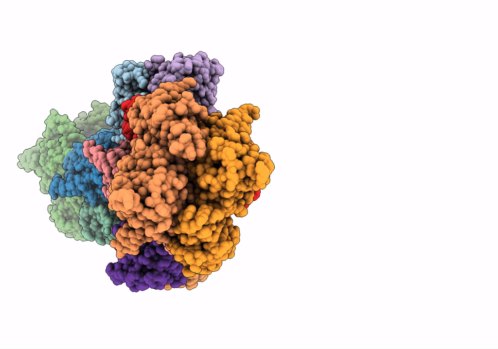
Biological Source:
Source Organism(s):
Escherichia coli (strain K12) (Taxon ID: 83333)
Escherichia virus T3 (Taxon ID: 2732706)
Escherichia virus T3 (Taxon ID: 2732706)
Expression System(s):
Method Details:
Experimental Method:
Resolution:
3.60 Å
Aggregation State:
PARTICLE
Reconstruction Method:
SINGLE PARTICLE