7OC8 image
Entry Detail
PDB ID:
7OC8
Keywords:
Title:
Trichoderma reesei Cel7A E212Q mutant in complex with pNPL
Biological Source:
Source Organism:
PDB Version:
Deposition Date:
2021-04-26
Release Date:
2022-03-09
Method Details:
Experimental Method:
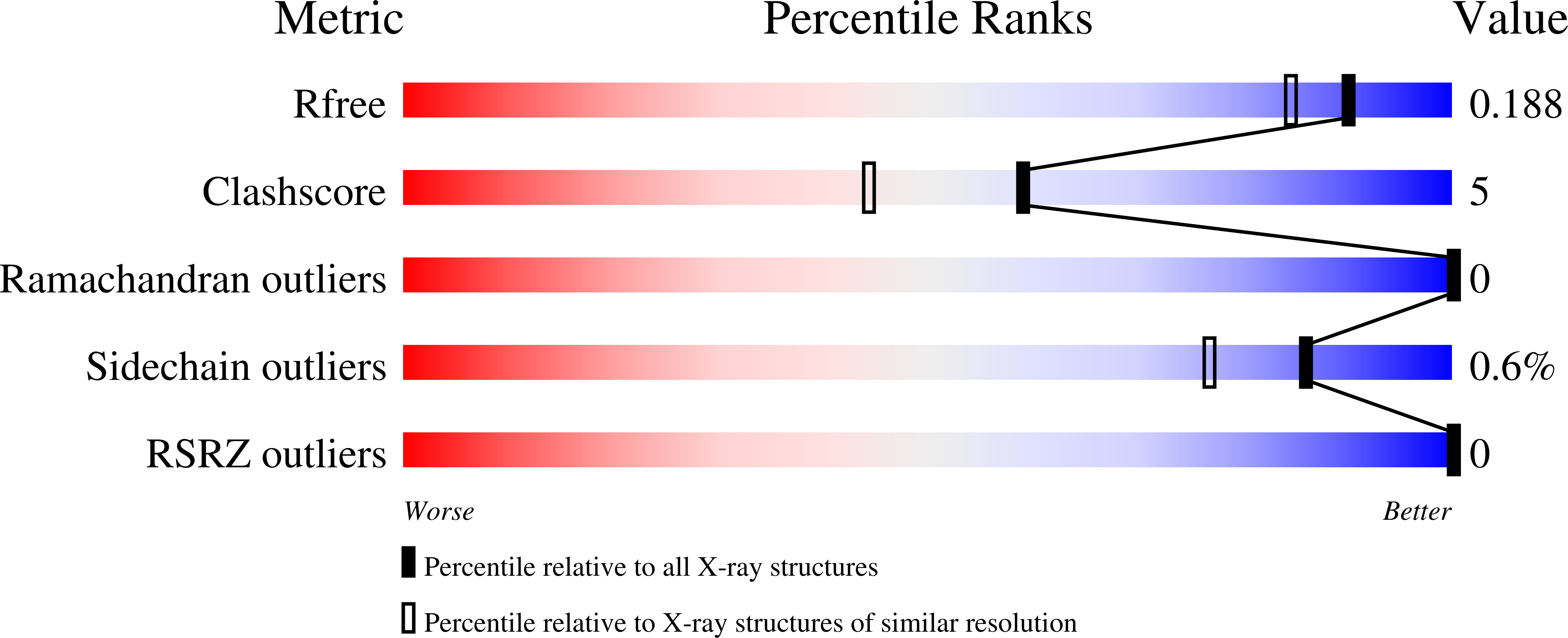
Resolution:
1.60 Å
R-Value Free:
0.18
R-Value Work:
0.15
R-Value Observed:
0.15
Space Group:
I 2 2 2
Macromolecular Entities
Polymer Type:polypeptide(L)
Description:Exoglucanase 1
Mutations:E212Q
Chain IDs:A
Chain Length:434
Number of Molecules:1
Biological Source:Hypocrea jecorina
Modified Residue
Compound ID Chain ID Parent Comp ID Details 2D Image
PCA A GLN modified residue
Primary Citation
Enzyme kinetics by GH7 cellobiohydrolases on chromogenic substrates is dictated by non-productive binding: insights from crystal structures and MD simulation.
Febs J. 290 379 399 (2023)
PMID: 35997626 DOI: 10.1111/febs.16602

Abstact

Cellobiohydrolases (CBHs) in the glycoside hydrolase family 7 (GH7) (EC3.2.1.176) are the major cellulose degrading enzymes both in industrial settings and in the context of carbon cycling in nature. Small carbohydrate conjugates such as p-nitrophenyl-β-d-cellobioside (pNPC), p-nitrophenyl-β-d-lactoside (pNPL) and methylumbelliferyl-β-d-cellobioside have commonly been used in colorimetric and fluorometric assays for analysing activity of these enzymes. Despite the similar nature of these compounds the kinetics of their enzymatic hydrolysis vary greatly between the different compounds as well as among different enzymes within the GH7 family. Through enzyme kinetics, crystallographic structure determination, molecular dynamics simulations, and fluorometric binding studies using the closely related compound o-nitrophenyl-β-d-cellobioside (oNPC), in this work we examine the different hydrolysis characteristics of these compounds on two model enzymes of this class, TrCel7A from Trichoderma reesei and PcCel7D from Phanerochaete chrysosporium. Protein crystal structures of the E212Q mutant of TrCel7A with pNPC and pNPL, and the wildtype TrCel7A with oNPC, reveal that non-productive binding at the product site is the dominating binding mode for these compounds. Enzyme kinetics results suggest the strength of non-productive binding is a key determinant for the activity characteristics on these substrates, with PcCel7D consistently showing higher turnover rates (kcat) than TrCel7A, but higher Michaelis-Menten (KM) constants as well. Furthermore, oNPC turned out to be useful as an active-site probe for fluorometric determination of the dissociation constant for cellobiose on TrCel7A but could not be utilized for the same purpose on PcCel7D, likely due to strong binding to an unknown site outside the active site.

Legend

Protein

Chemical

Disease

Primary Citation of related structures