7O6A image
Deposition Date 2021-04-09
Release Date 2021-10-13
Last Version Date 2024-01-31
Entry Detail
PDB ID:
7O6A
Keywords:
Title:
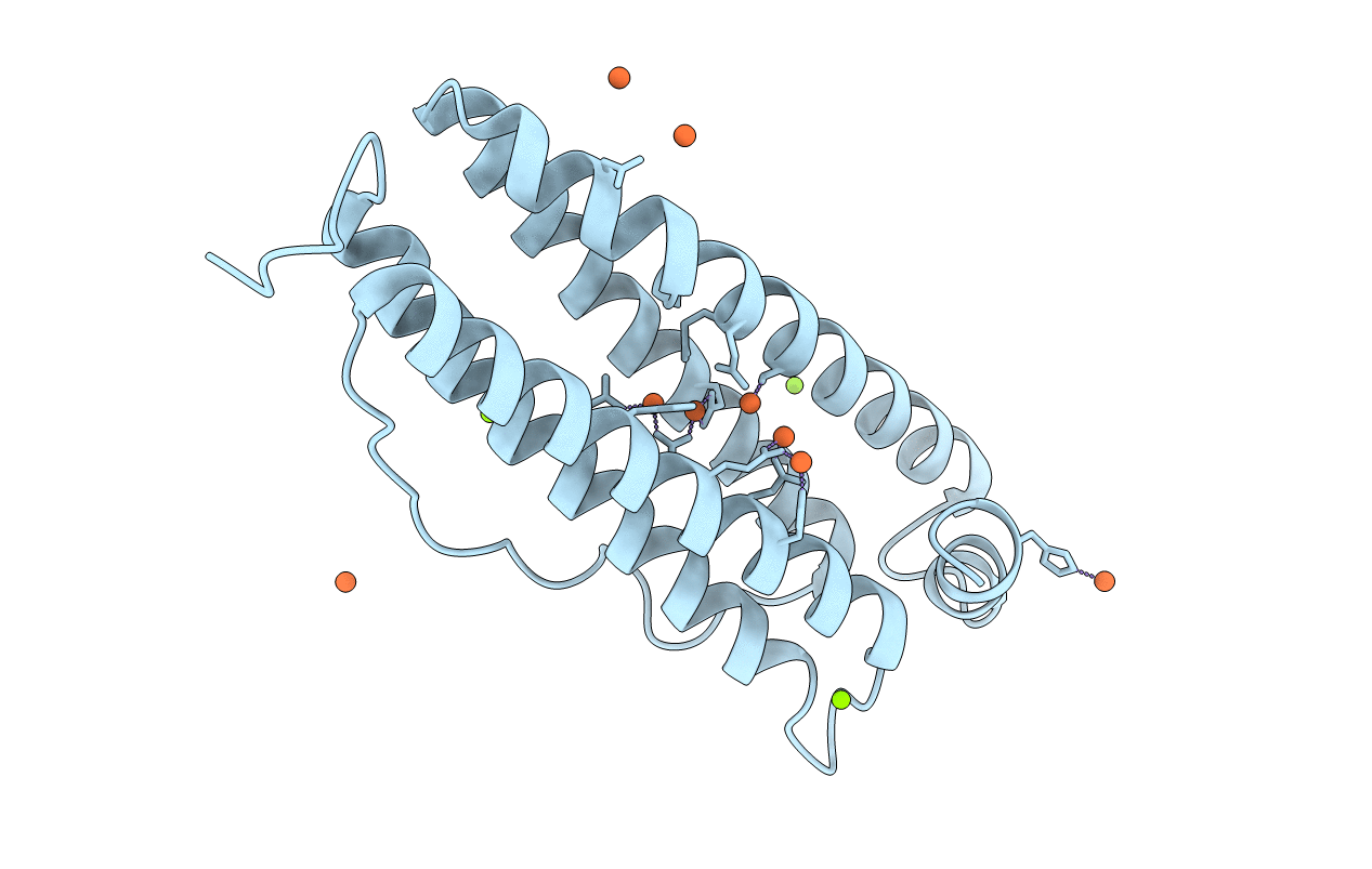
Crystal structure of human mitochondrial ferritin (hMTF) Fe(II)-loaded for 5 minutes under anaerobic environment
Biological Source:
Source Organism:
Homo sapiens (Taxon ID: 9606)
Method Details:
Experimental Method:
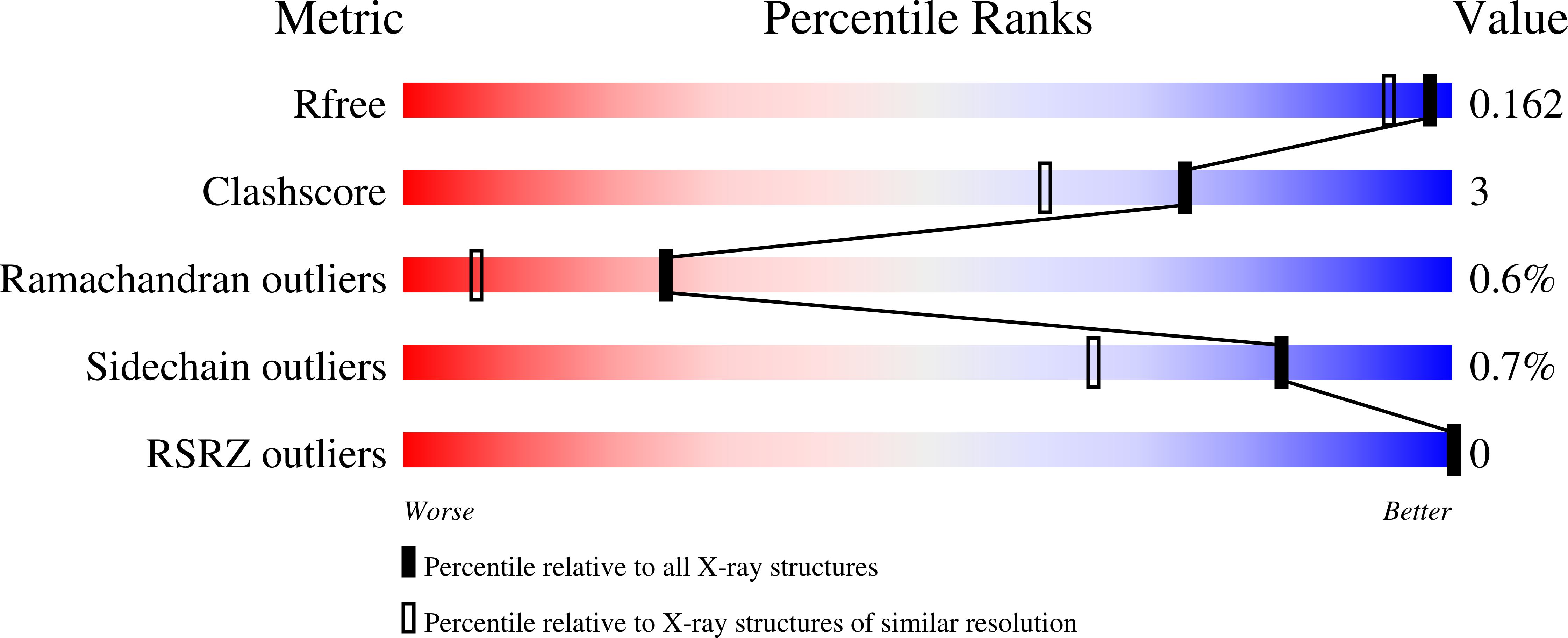
Resolution:
1.40 Å
R-Value Free:
0.16
R-Value Work:
0.13
R-Value Observed:
0.13
Space Group:
F 4 3 2
Macromolecular Entities
Polymer Type:polypeptide(L)
Molecule:Ferritin, mitochondrial
Gene (Uniprot):FTMT
Chain IDs:A
Chain Length:183
Number of Molecules:1
Biological Source:Homo sapiens
Primary Citation
Iron Binding in the Ferroxidase Site of Human Mitochondrial Ferritin.
Chemistry 27 14690 14701 (2021)
PMID: 34343376 DOI: 10.1002/chem.202102270

Abstact

Ferritins are nanocage proteins that store iron ions in their central cavity as hydrated ferric oxide biominerals. In mammals, further the L (light) and H (heavy) chains constituting cytoplasmic maxi-ferritins, an additional type of ferritin has been identified, the mitochondrial ferritin (MTF). Human MTF (hMTF) is a functional homopolymeric H-like ferritin performing the ferroxidase activity in its ferroxidase site (FS), in which Fe(II) is oxidized to Fe(III) in the presence of dioxygen. To better investigate its ferroxidase properties, here we performed time-lapse X-ray crystallography analysis of hMTF, providing structural evidence of how iron ions interact with hMTF and of their binding to the FS. Transient iron binding sites, populating the pathway along the cage from the iron entry channel to the catalytic center, were also identified. Furthermore, our kinetic data at variable iron loads indicate that the catalytic iron oxidation reaction occurs via a diferric peroxo intermediate followed by the formation of ferric-oxo species, with significant differences with respect to human H-type ferritin.

Legend

Protein

Chemical

Disease

Primary Citation of related structures