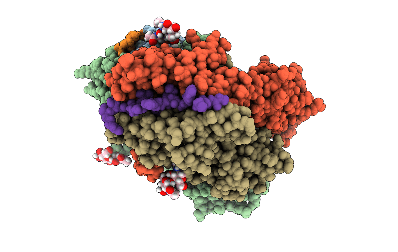
7NZH image
Entry Detail
PDB ID:
7NZH
Keywords:
Title:
Crystal structure of HLA-DR4 in complex with a citrullinated cilp peptide
Biological Source:
Source Organism:
Host Organism:
PDB Version:
Deposition Date:
2021-03-24
Release Date:
2022-05-11
Method Details:
Experimental Method:
Resolution:
2.83 Å
R-Value Free:
0.28
R-Value Work:
0.24
Space Group:
C 2 2 21
Macromolecular Entities
Polymer Type:polypeptide(L)
Description:HLA class II histocompatibility antigen, DR alpha chain
Chain IDs:A (auth: AAA), C (auth: CCC)
Chain Length:180
Number of Molecules:2
Biological Source:Homo sapiens
Polymer Type:polypeptide(L)
Description:HLA class II histocompatibility antigen, DR beta chain
Chain IDs:B (auth: BBB), D (auth: DDD)
Chain Length:189
Number of Molecules:2
Biological Source:Homo sapiens
Polymer Type:polypeptide(L)
Description:citrullinated cartilage intermediate layer protein (CILP) peptide 982-996
Chain IDs:E (auth: EEE), F (auth: FFF)
Chain Length:14
Number of Molecules:2
Biological Source:Homo sapiens
Ligand Molecules
Primary Citation
Key interactions in the trimolecular complex consisting of the rheumatoid arthritis-associated DRB1*04:01 molecule, the major glycosylated collagen II peptide and the T-cell receptor.
Ann Rheum Dis 81 480 489 (2022)
PMID: 35027402 DOI: 10.1136/annrheumdis-2021-220500

Abstact

OBJECTIVES Rheumatoid arthritis (RA) is an autoimmune disease strongly associated with the major histocompatibility complex (MHC) class II allele DRB1*04:01, which encodes a protein that binds self-peptides for presentation to T cells. This study characterises the autoantigen-presenting function of DRB1*04:01 (HLA-DRA*01:01/HLA-DRB1*04:01) at a molecular level for prototypic T-cell determinants, focusing on a post-translationally modified collagen type II (Col2)-derived peptide. METHODS The crystal structures of DRB1*04:01 molecules in complex with the peptides HSP70289-306, citrullinated CILP982-996 and galactosylated Col2259-273 were determined on cocrystallisation. T cells specific for Col2259-273 were investigated in peripheral blood mononuclear cells from patients with DRB1*04:01-positive RA by cytofluorometric detection of the activation marker CD154 on peptide stimulation and binding of fluorescent DRB1*0401/Col2259-273 tetramer complexes. The cDNAs encoding the T-cell receptor (TCR) α-chains and β-chains were cloned from single-cell sorted tetramer-positive T cells and transferred via a lentiviral vector into TCR-deficient Jurkat 76 cells. RESULTS The crystal structures identified peptide binding to DRB1*04:01 and potential side chain exposure to T cells. The main TCR recognition sites in Col2259-273 were lysine residues that can be galactosylated. RA T-cell responses to DRB1*04:01-presented Col2259-273 were dependent on peptide galactosylation at lysine 264. Dynamic molecular modelling of a functionally characterised Col2259-273-specific TCR complexed with DRB1*04:01/Col2259-273 provided evidence for differential allosteric T-cell recognition of glycosylated lysine 264. CONCLUSIONS The MHC-peptide-TCR interactions elucidated in our study provide new molecular insights into recognition of a post-translationally modified RA T-cell determinant with a known dominant role in arthritogenic and tolerogenic responses in murine Col2-induced arthritis.

Legend

Protein

Chemical

Disease

Primary Citation of related structures