7NXL image
Deposition Date 2021-03-18
Release Date 2021-09-08
Last Version Date 2024-11-06
Entry Detail
PDB ID:
7NXL
Keywords:
Title:
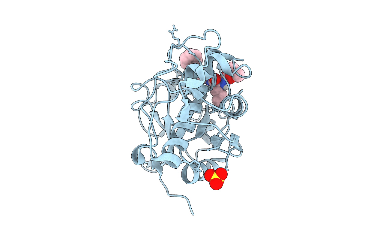
Structure of human cathepsin K in complex with the acrylamide inhibitor Gu3110
Biological Source:
Source Organism:
Homo sapiens (Taxon ID: 9606)
Host Organism:
Method Details:
Experimental Method:
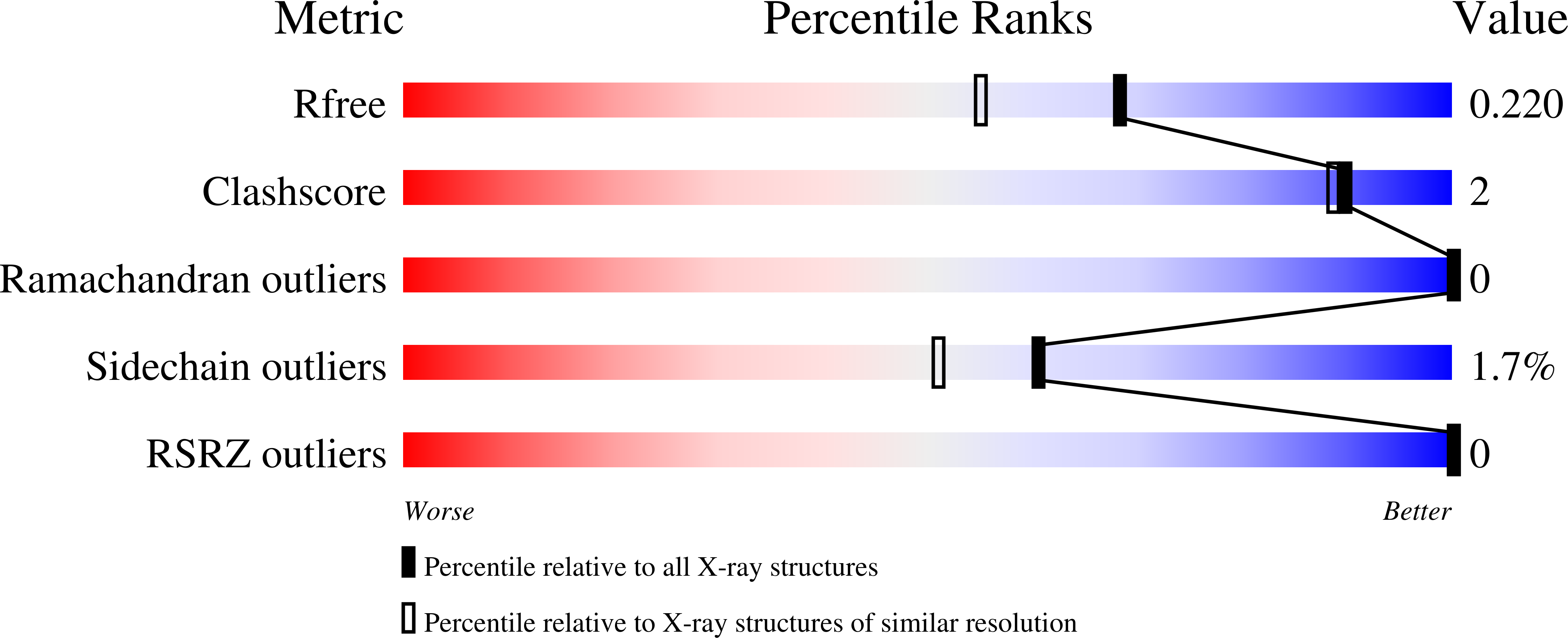
Resolution:
1.80 Å
R-Value Free:
0.20
R-Value Work:
0.17
R-Value Observed:
0.17
Space Group:
P 2 21 21
Macromolecular Entities
Polymer Type:polypeptide(L)
Molecule:Cathepsin K
Gene (Uniprot):CTSK
Chain IDs:A (auth: AAA)
Chain Length:216
Number of Molecules:1
Biological Source:Homo sapiens
Primary Citation
An Activity-Based Probe for Cathepsin K Imaging with Excellent Potency and Selectivity.
J.Med.Chem. 64 13793 13806 (2021)
PMID: 34473502 DOI: 10.1021/acs.jmedchem.1c01178

Abstact

The cysteine protease cathepsin K is a target for the treatment of diseases associated with high bone turnover. Cathepsin K is mainly expressed in osteoclasts and responsible for the destruction of the proteinaceous components of the bone matrix. We designed various fluorescent activity-based probes (ABPs) and their precursors that bind to and inactivate cathepsin K. ABP 25 exhibited extraordinary potency (kinac/Ki = 35,300 M-1s-1) and selectivity for human cathepsin K. Crystal structures of cathepsin K in complex with ABP 25 and its nonfluorescent precursor 21 were determined to characterize the binding mode of this new type of acrylamide-based Michael acceptor with the particular orientation of the dibenzylamine moiety to the primed subsite region. The cyanine-5 containing probe 25 allowed for sensitive detection of cathepsin K, selective visualization in complex proteomes, and live cell imaging of a human osteosarcoma cell line, underlining its applicability in a pathophysiological environment.

Legend

Protein

Chemical

Disease

Primary Citation of related structures