7NWO image
Entry Detail
PDB ID:
7NWO
Title:
A carbohydrate binding module family 9 (CBM9) from Caldicellulosiruptor kristjanssonii in complex with glucose
Biological Source:
PDB Version:
Deposition Date:
2021-03-17
Release Date:
2021-07-07
Method Details:
Experimental Method:
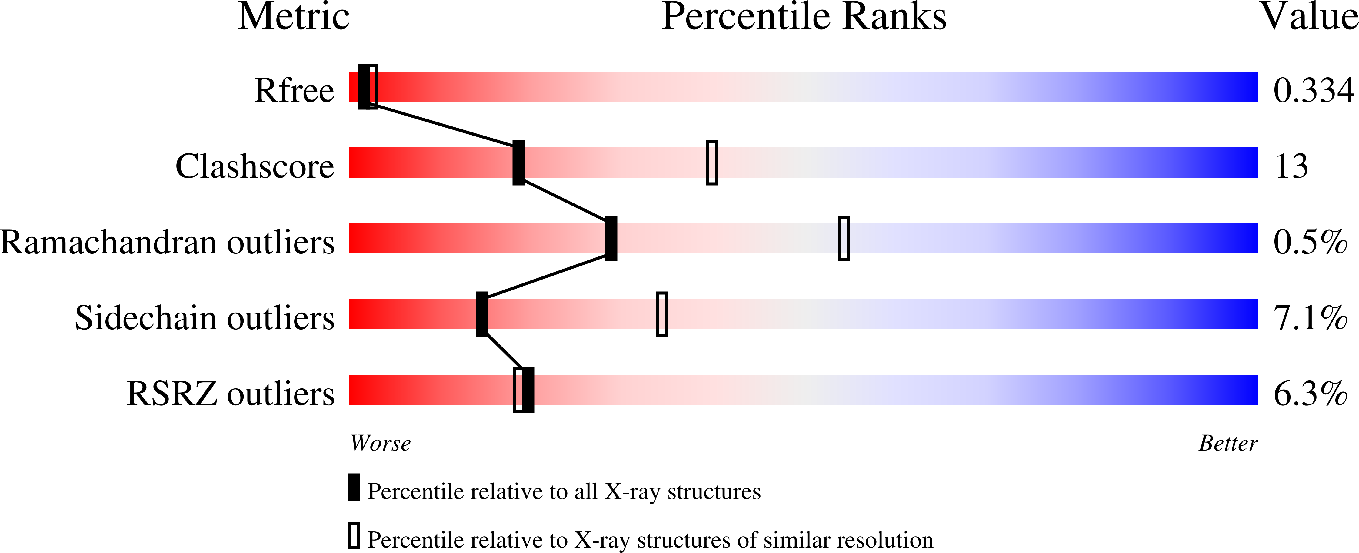
Resolution:
2.70 Å
R-Value Free:
0.33
R-Value Work:
0.25
Space Group:
I 4 3 2
Macromolecular Entities
Polymer Type:polypeptide(L)
Description:Beta-xylanase
Chain IDs:A (auth: AAA)
Chain Length:215
Number of Molecules:1
Biological Source:Caldicellulosiruptor kristjanssonii (strain ATCC 700853 / DSM 12137 / I77R1B)
Primary Citation
Structural and Functional Analysis of a Multimodular Hyperthermostable Xylanase-Glucuronoyl Esterase from Caldicellulosiruptor kristjansonii .
Biochemistry 60 2206 2220 (2021)
PMID: 34180241 DOI: 10.1021/acs.biochem.1c00305

Abstact

The hyperthermophilic bacterium Caldicellulosiruptor kristjansonii encodes an unusual enzyme, CkXyn10C-GE15A, which incorporates two catalytic domains, a xylanase and a glucuronoyl esterase, and five carbohydrate-binding modules (CBMs) from families 9 and 22. The xylanase and glucuronoyl esterase catalytic domains were recently biochemically characterized, as was the ability of the individual CBMs to bind insoluble polysaccharides. Here, we further probed the abilities of the different CBMs from CkXyn10C-GE15A to bind to soluble poly- and oligosaccharides using affinity gel electrophoresis, isothermal titration calorimetry, and differential scanning fluorimetry. The results revealed additional binding properties of the proteins compared to the former studies on insoluble polysaccharides. Collectively, the results show that all five CBMs have their own distinct binding preferences and appear to complement each other and the catalytic domains in targeting complex cell wall polysaccharides. Additionally, through renewed efforts, we have achieved partial structural characterization of this complex multidomain protein. We have determined the structures of the third CBM9 domain (CBM9.3) and the glucuronoyl esterase (GE15A) by X-ray crystallography. CBM9.3 is the second CBM9 structure determined to date and was shown to bind oligosaccharide ligands at the same site but in a different binding mode compared to that of the previously determined CBM9 structure from Thermotoga maritima. GE15A represents a unique intermediate between reported fungal and bacterial glucuronoyl esterase structures as it lacks two inserted loop regions typical of bacterial enzymes and a third loop has an atypical structure. We also report small-angle X-ray scattering measurements of the N-terminal CBM22.1-CBM22.2-Xyn10C construct, indicating a compact arrangement at room temperature.

Legend

Protein

Chemical

Disease

Primary Citation of related structures