7NSL image
Deposition Date 2021-03-08
Release Date 2021-11-24
Last Version Date 2024-10-23
Entry Detail
PDB ID:
7NSL
Keywords:
Title:
AL amyloid fibril from a lambda 1 light chain
Biological Source:
Source Organism:
Homo sapiens (Taxon ID: 9606)
Method Details:
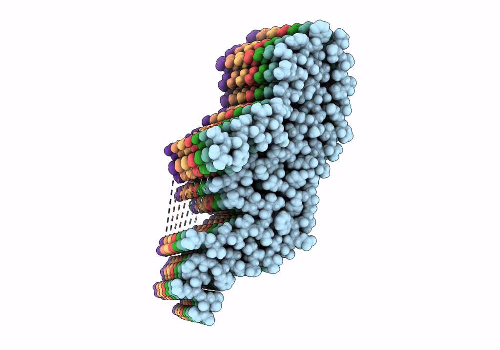
Experimental Method:
Resolution:
3.10 Å
Aggregation State:
FILAMENT
Reconstruction Method:
HELICAL
Macromolecular Entities
Polymer Type:polypeptide(L)
Molecule:Amyloid lambda1 light chain
Chain IDs:A (auth: D), B (auth: E), C (auth: F), D (auth: C), E (auth: B), F (auth: A), G
Chain Length:118
Number of Molecules:7
Biological Source:Homo sapiens
Ligand Molecules
Primary Citation
Role of mutations and post-translational modifications in systemic AL amyloidosis studied by cryo-EM.
Nat Commun 12 6434 6434 (2021)
PMID: 34741031 DOI: 10.1038/s41467-021-26553-9

Abstact

Systemic AL amyloidosis is a rare disease that is caused by the misfolding of immunoglobulin light chains (LCs). Potential drivers of amyloid formation in this disease are post-translational modifications (PTMs) and the mutational changes that are inserted into the LCs by somatic hypermutation. Here we present the cryo electron microscopy (cryo-EM) structure of an ex vivo λ1-AL amyloid fibril whose deposits disrupt the ordered cardiomyocyte structure in the heart. The fibril protein contains six mutational changes compared to the germ line and three PTMs (disulfide bond, N-glycosylation and pyroglutamylation). Our data imply that the disulfide bond, glycosylation and mutational changes contribute to determining the fibril protein fold and help to generate a fibril morphology that is able to withstand proteolytic degradation inside the body.

Legend

Protein

Chemical

Disease

Primary Citation of related structures