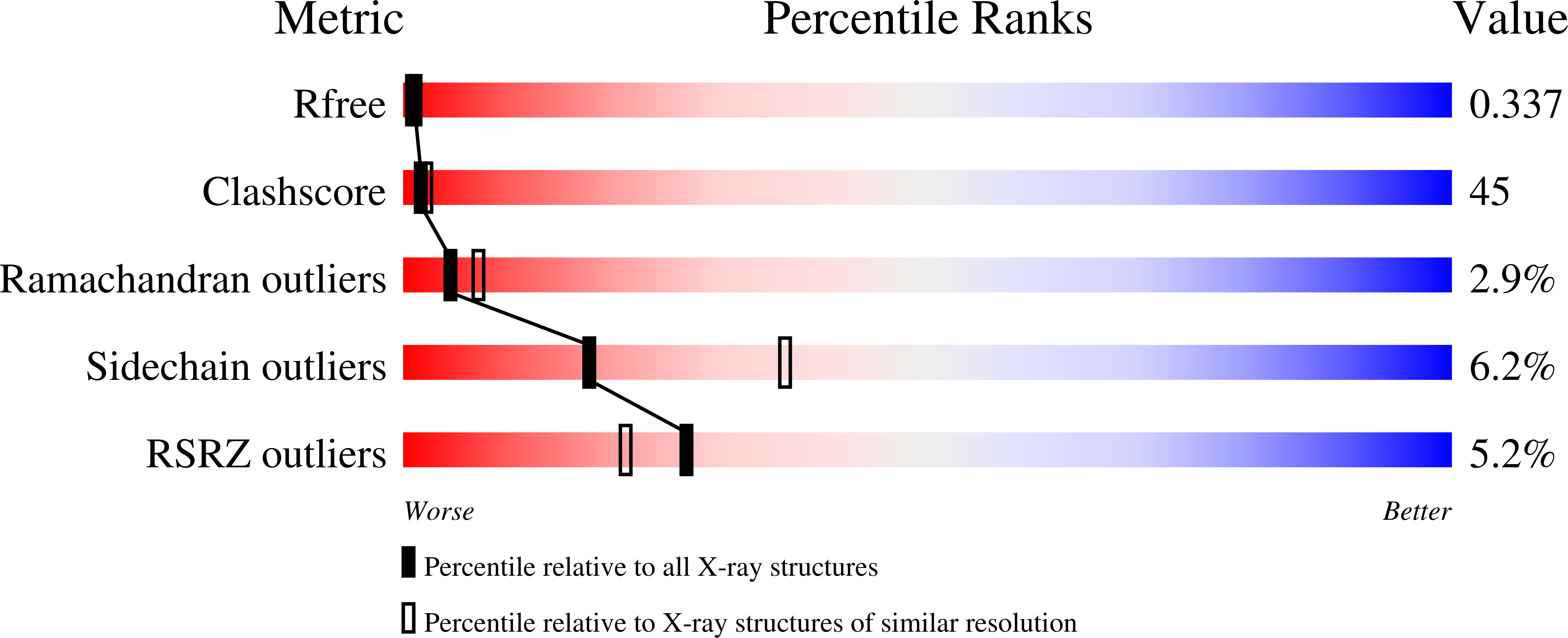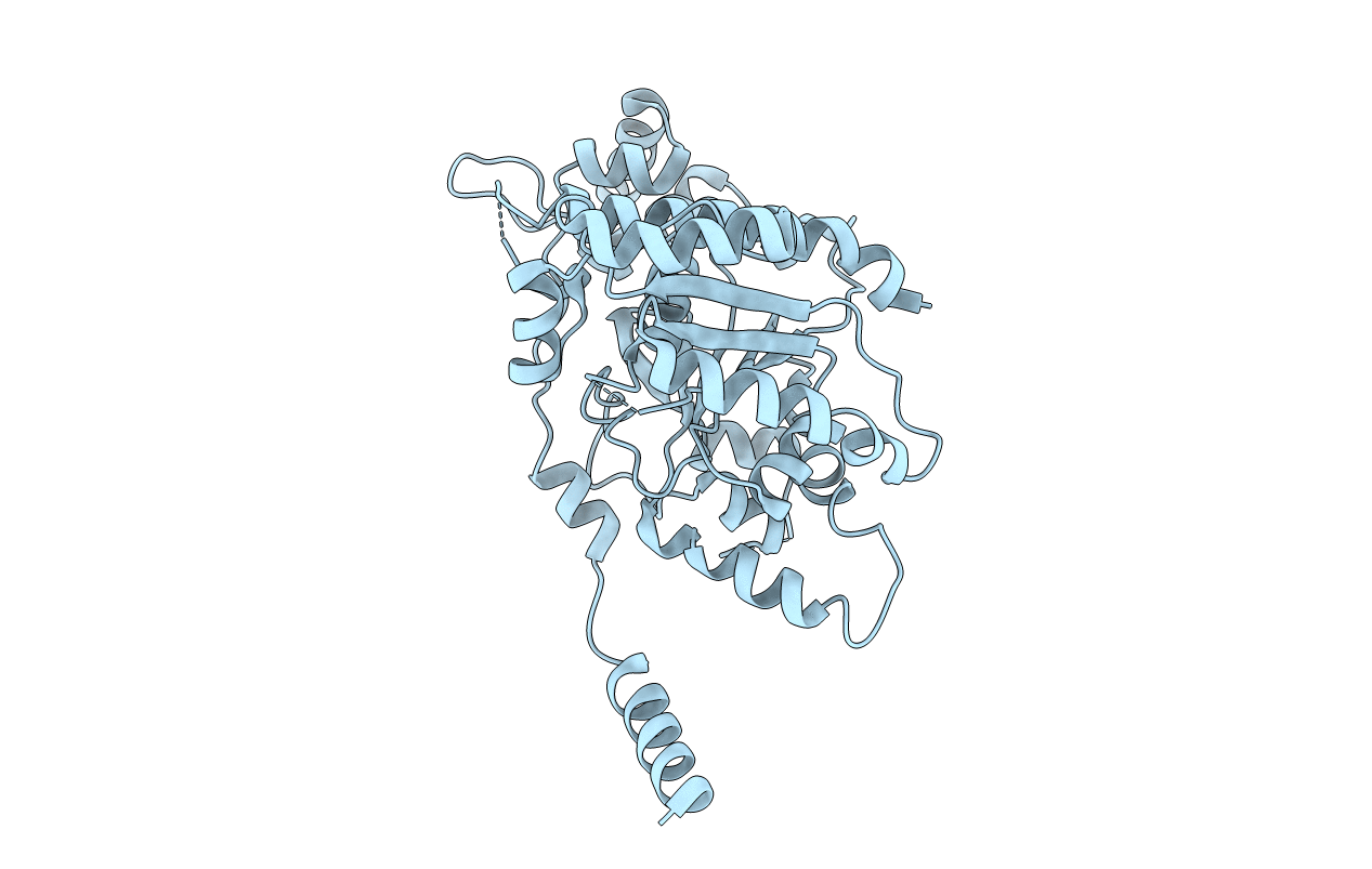
7NPD image
Deposition Date 2021-02-26
Release Date 2021-05-19
Last Version Date 2024-01-31
Entry Detail
PDB ID:
7NPD
Title:
Vibiro cholerae ParA2
Biological Source:
Source Organism:
Vibrio cholerae (Taxon ID: 666)
Host Organism:
Method Details:
Experimental Method:
Resolution:
2.60 Å
R-Value Free:
0.33
R-Value Work:
0.27
R-Value Observed:
0.28
Space Group:
P 32 1 2
Macromolecular Entities
Polymer Type:polypeptide(L)
Molecule:Walker A-type ATPase
Chain IDs:A
Chain Length:407
Number of Molecules:1
Biological Source:Vibrio cholerae
Primary Citation
The structure of the bacterial DNA segregation ATPase filament reveals the conformational plasticity of ParA upon DNA binding.
Nat Commun 12 5166 5166 (2021)
PMID: 34453062 DOI: 10.1038/s41467-021-25429-2

Abstact

The efficient segregation of replicated genetic material is an essential step for cell division. Bacterial cells use several evolutionarily-distinct genome segregation systems, the most common of which is the type I Par system. It consists of an adapter protein, ParB, that binds to the DNA cargo via interaction with the parS DNA sequence; and an ATPase, ParA, that binds nonspecific DNA and mediates cargo transport. However, the molecular details of how this system functions are not well understood. Here, we report the cryo-EM structure of the Vibrio cholerae ParA2 filament bound to DNA, as well as the crystal structures of this protein in various nucleotide states. These structures show that ParA forms a left-handed filament on DNA, stabilized by nucleotide binding, and that ParA undergoes profound structural rearrangements upon DNA binding and filament assembly. Collectively, our data suggest the structural basis for ParA's cooperative binding to DNA and the formation of high ParA density regions on the nucleoid.

Legend

Protein

Chemical

Disease

Primary Citation of related structures