7N6B image
Deposition Date 2021-06-08
Release Date 2021-09-01
Last Version Date 2025-05-21
Entry Detail
PDB ID:
7N6B
Keywords:
Title:
Structure of MmpL3 reconstituted into lipid nanodisc in the TMM bound state
Biological Source:
Method Details:
Experimental Method:
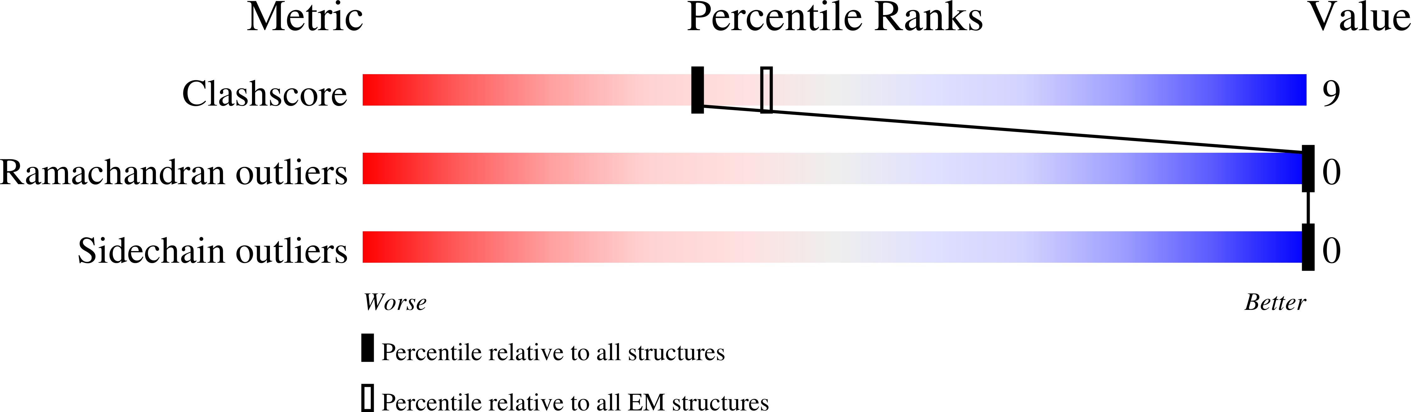
Resolution:
2.66 Å
Aggregation State:
PARTICLE
Reconstruction Method:
SINGLE PARTICLE
Macromolecular Entities
Structures with similar UniProt ID
Protein Blast
Polymer Type:polypeptide(L)
Molecule:MmpL3 transporter
Chain IDs:A
Chain Length:1013
Number of Molecules:1
Biological Source:Mycolicibacterium smegmatis (strain ATCC 700084 / mc(2)155)
Ligand Molecules
Primary Citation
Structures of the mycobacterial membrane protein MmpL3 reveal its mechanism of lipid transport.
Plos Biol. 19 e3001370 e3001370 (2021)
PMID: 34383749 DOI: 10.1371/journal.pbio.3001370

Abstact

The mycobacterial membrane protein large 3 (MmpL3) transporter is essential and required for shuttling the lipid trehalose monomycolate (TMM), a precursor of mycolic acid (MA)-containing trehalose dimycolate (TDM) and mycolyl arabinogalactan peptidoglycan (mAGP), in Mycobacterium species, including Mycobacterium tuberculosis and Mycobacterium smegmatis. However, the mechanism that MmpL3 uses to facilitate the transport of fatty acids and lipidic elements to the mycobacterial cell wall remains elusive. Here, we report 7 structures of the M. smegmatis MmpL3 transporter in its unbound state and in complex with trehalose 6-decanoate (T6D) or TMM using single-particle cryo-electron microscopy (cryo-EM) and X-ray crystallography. Combined with calculated results from molecular dynamics (MD) and target MD simulations, we reveal a lipid transport mechanism that involves a coupled movement of the periplasmic domain and transmembrane helices of the MmpL3 transporter that facilitates the shuttling of lipids to the mycobacterial cell wall.

Legend

Protein

Chemical

Disease

Primary Citation of related structures