
Deposition Date
2021-05-24
Release Date
2021-06-02
Last Version Date
2023-10-18
Entry Detail
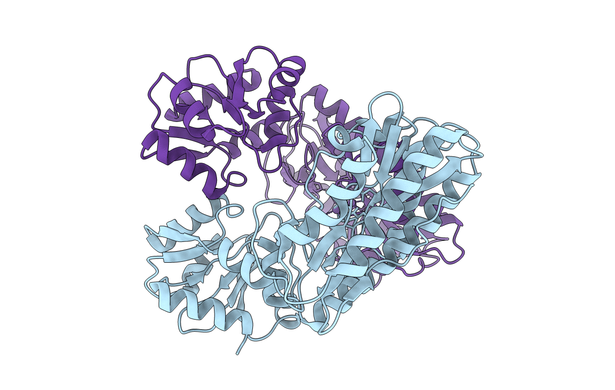
PDB ID:
7N02
Keywords:
Title:
X-ray crystallographic structure model of Lactococcus lactis prolidase mutant D36S
Biological Source:
Source Organism(s):
Lactococcus lactis (Taxon ID: 1358)
Expression System(s):
Method Details:
Experimental Method:
Resolution:
2.35 Å
R-Value Free:
0.26
R-Value Work:
0.21
R-Value Observed:
0.21
Space Group:
P 21 21 21
